Method for positioning the breast for a biopsy in a mammography device, and mammography device to implement the method

a mammography and breast technology, applied in the field of breast biopsy positioning in a mammography device, to achieve the effect of reducing the number of x-ray images required for correct positioning

Active Publication Date: 2010-03-04
SIEMENS HEALTHCARE GMBH
View PDF7 Cites 28 Cited by
  • Summary
  • Abstract
  • Description
  • Claims
  • Application Information

AI Technical Summary

Benefits of technology

[0005]An object of the invention is to provide a method for positioning the breast for a biopsy in a mammography apparatus with which the number of the x-ray

Problems solved by technology

However, every such x-ray acquisition entails a

Method used


Image

  • Method for positioning the breast for a biopsy in a mammography device, and mammography device to implement the method
  • Method for positioning the breast for a biopsy in a mammography device, and mammography device to implement the method
  • Method for positioning the breast for a biopsy in a mammography device, and mammography device to implement the method

Examples


Embodiment Construction

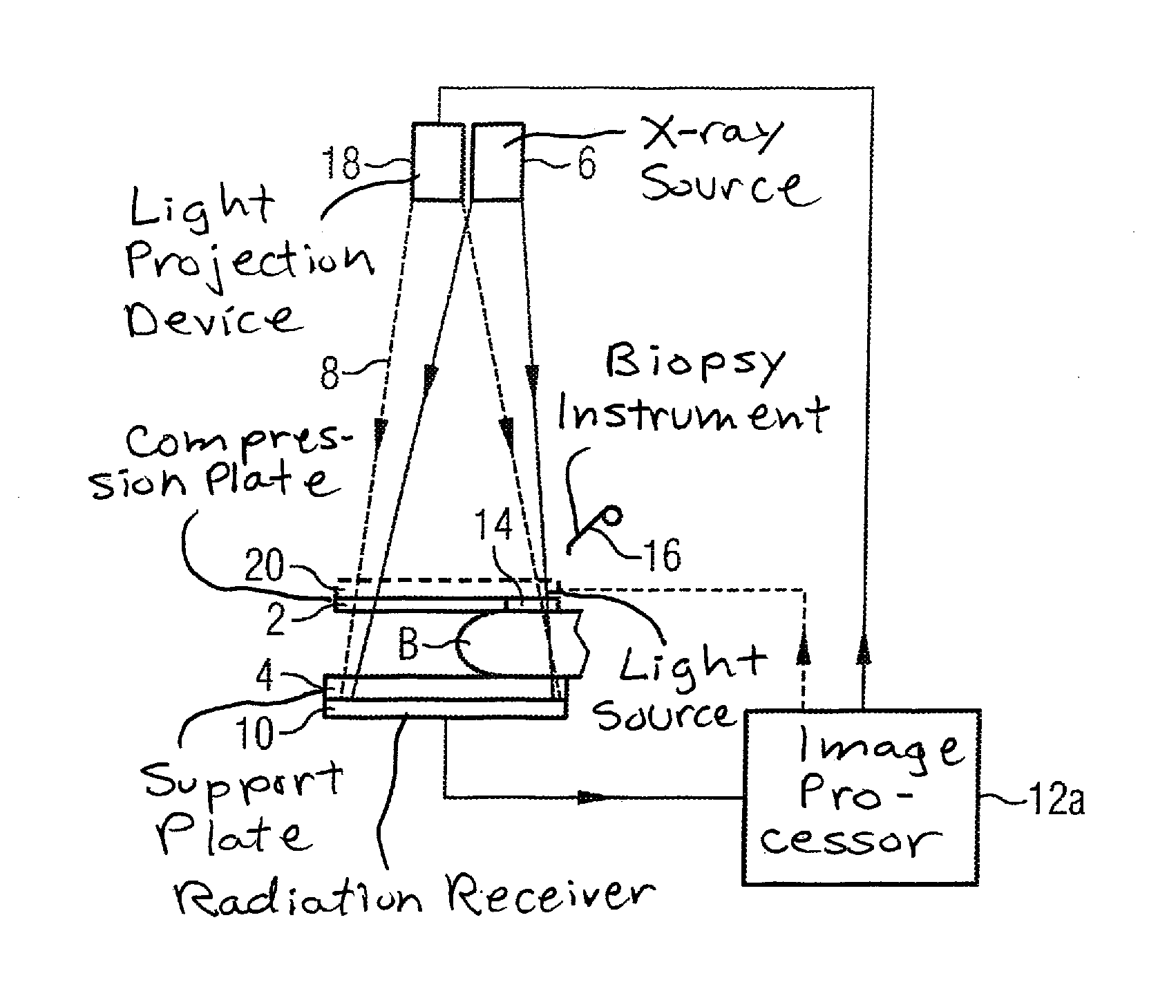
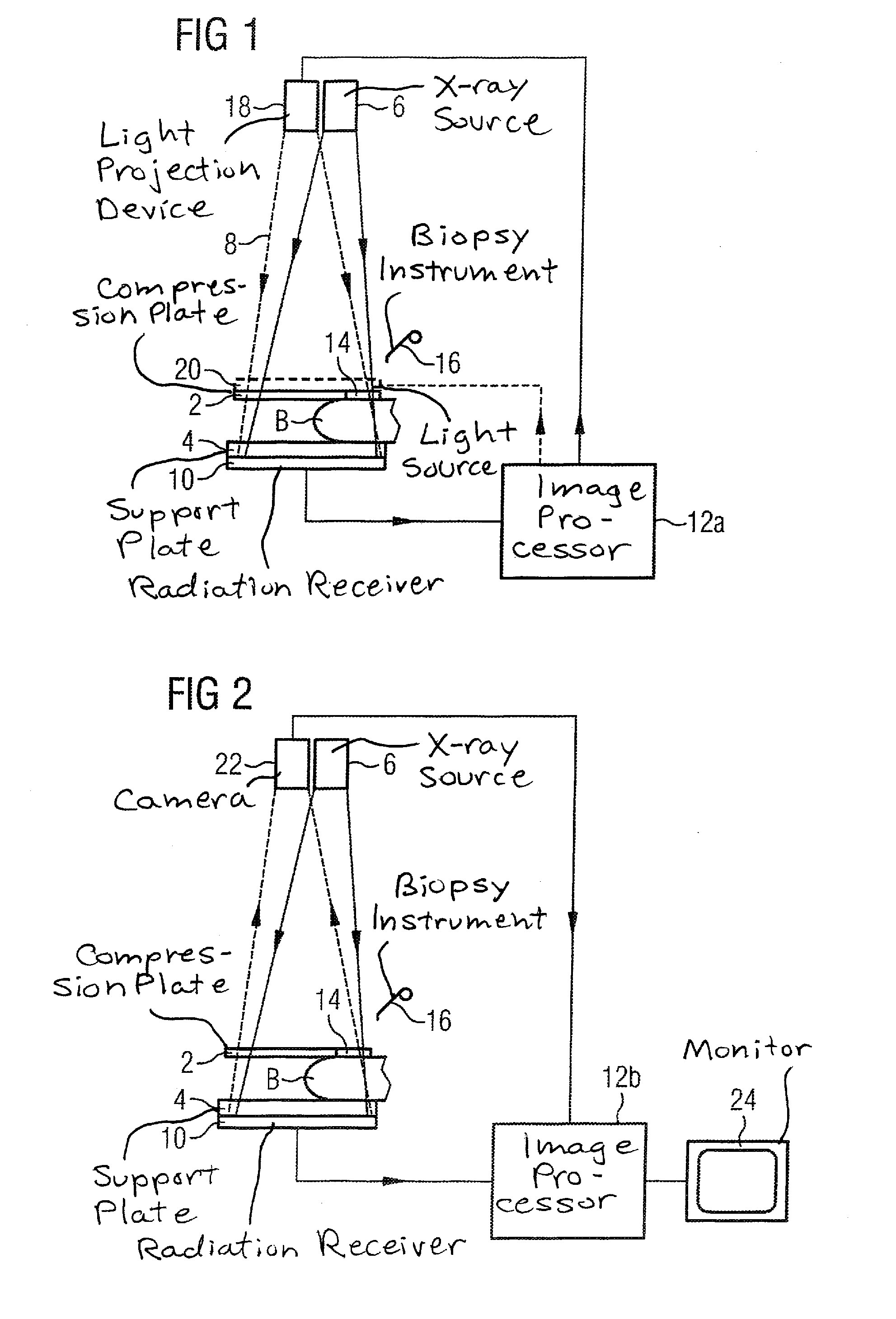
[0026]According to FIG. 1, a breast B is compressed and fixed between a compression plate 2 and a support plate 4 in a mammography device according to the invention. To generate an x-ray image of the breast B, x-rays 8 are generated by an x-ray source 6 and received (detected) by an x-ray receiver 10 arranged after the bearing plate 4. The image data acquired by the x-ray receiver 10 are processed in an image processor 12a.

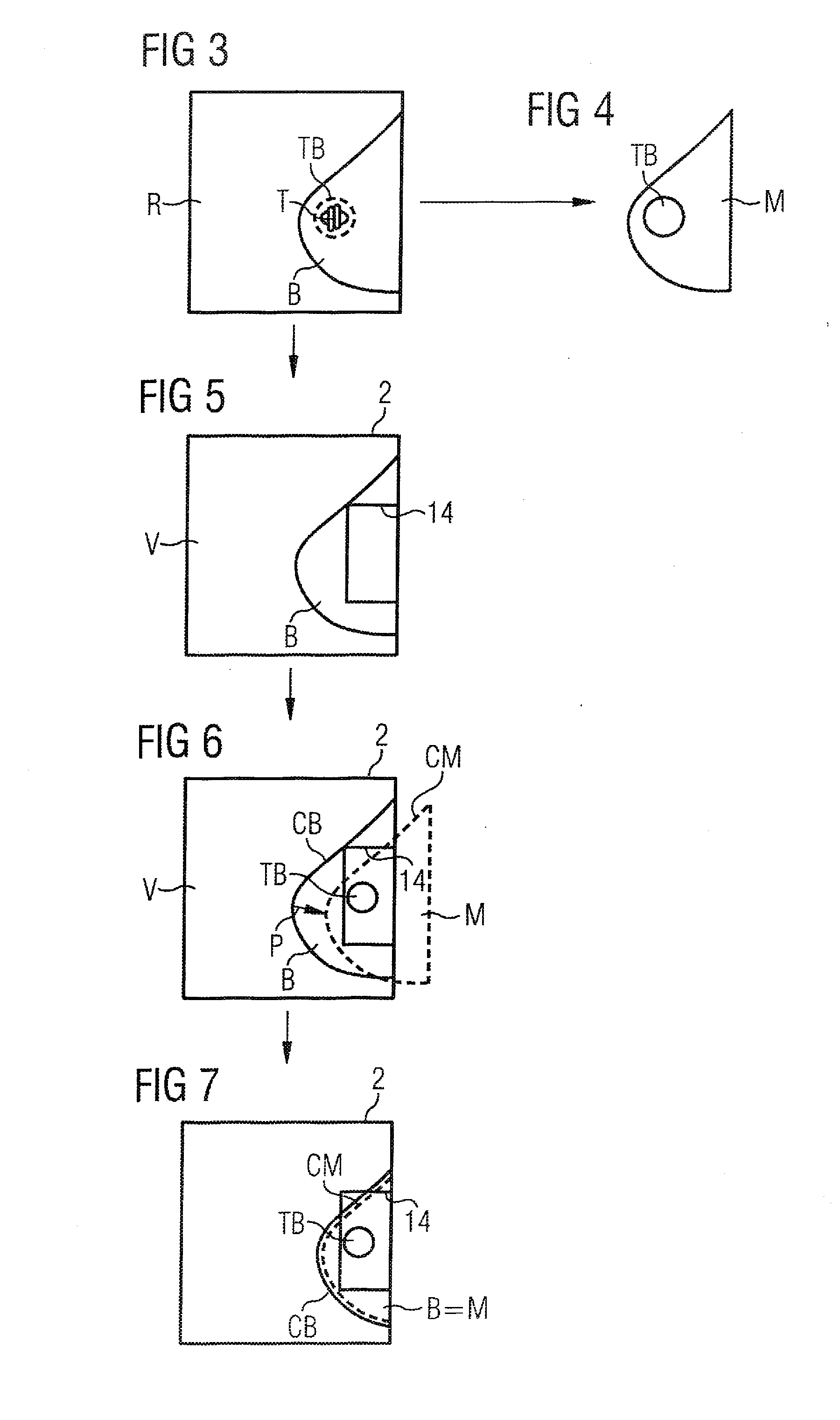
[0027]The compression plate 2 has a rectangular opening 14 through which a biopsy instrument 16 can be inserted into the breast B. An x-ray image of the breast B that has been generated and assessed either shortly beforehand with the same mammography device or at a point in time further in the past with a different mammography device is located in the memory of the image processor 12a. From this x-ray image a virtual mask is now generated in the image processor 12a that renders the image region covered by the breast tissue in the x-ray image, i.e. whose edge coin...


PUM


Abstract

In a method and a mammography device, a virtual mask is created from an x-ray image of the breast for positioning the breast for a biopsy. The mask reproduces the image region covered by the breast tissue. A region targeted for the biopsy, and located within the breast tissue, is marked in the mask. The breast is then positioned for the biopsy in the mammography device between a support plate and a compression plate, the compression plate being provided with a recess for introducing the biopsy instrument therethrough. The mask and the compression plate are superimposed to cause the marked region to be located within this recess. The breast is then moved until the position and the contour thereof substantially coincide with the position and contour of the mask.

Description

BACKGROUND OF THE INVENTION[0001]1. Field of the Invention[0002]The present invention concerns a method for positioning the breast for a biopsy in a mammography device. Moreover, the invention concerns a mammography device to implement this method.[0003]2. Description of the Prior Art[0004]When a pathological tissue variation or lesion is established in an x-ray image of the breast within the framework of a mammogram examination, for a definite diagnosis it is required in many cases to extract a tissue sample and to histologically examine this. For this purpose, in the stereotactic biopsy the breast is compressed and fixed between a compression plate and a bearing plate in a mammography device. The compression plate contains a rectangular opening via which the biopsy instrument can be inserted into the breast. In order to ensure that the tissue sample is extracted at the correct point, x-ray exposures are created from different directions during the procedure and the lesion is marke...

Claims


Application Information

Patent Timeline
IPC IPC(8): A61B6/04
CPCA61B6/08A61B6/542A61B6/502
Inventor FISCHER, DANIELHOFMANN, CARINAMERTELMEIER, THOMAS
Owner SIEMENS HEALTHCARE GMBH
Why Eureka
Try Eureka