Breast cancer pathological image diagnosis support system, breast cancer pathological image diagnosis support method, and recording medium recording breast cancer pathological image diagnosis support program

a breast cancer and pathological image technology, applied in diagnostic recording/measuring, instruments, applications, etc., can solve the problems of overwhelmingly small number of pathologists and inability to realize standardization of immunohistochemical staining examination methods and evaluation methods

Active Publication Date: 2010-03-04
NEC CORP
View PDF9 Cites 34 Cited by
  • Summary
  • Abstract
  • Description
  • Claims
  • Application Information

AI Technical Summary

Problems solved by technology

Although this pathological test is one of a test methods generally performed in current medical institutions, standardization of an examination method and an evaluation method of the immunohistochemical staining is not realized.
Although an automatic staining device for staining the tissue section is widely used and the number of cases of the pathological tests increases in these years, the number of pathologists is overwhelmingly small.

Method used


Image

  • Breast cancer pathological image diagnosis support system, breast cancer pathological image diagnosis support method, and recording medium recording breast cancer pathological image diagnosis support program
  • Breast cancer pathological image diagnosis support system, breast cancer pathological image diagnosis support method, and recording medium recording breast cancer pathological image diagnosis support program
  • Breast cancer pathological image diagnosis support system, breast cancer pathological image diagnosis support method, and recording medium recording breast cancer pathological image diagnosis support program

Examples


first exemplary embodiment

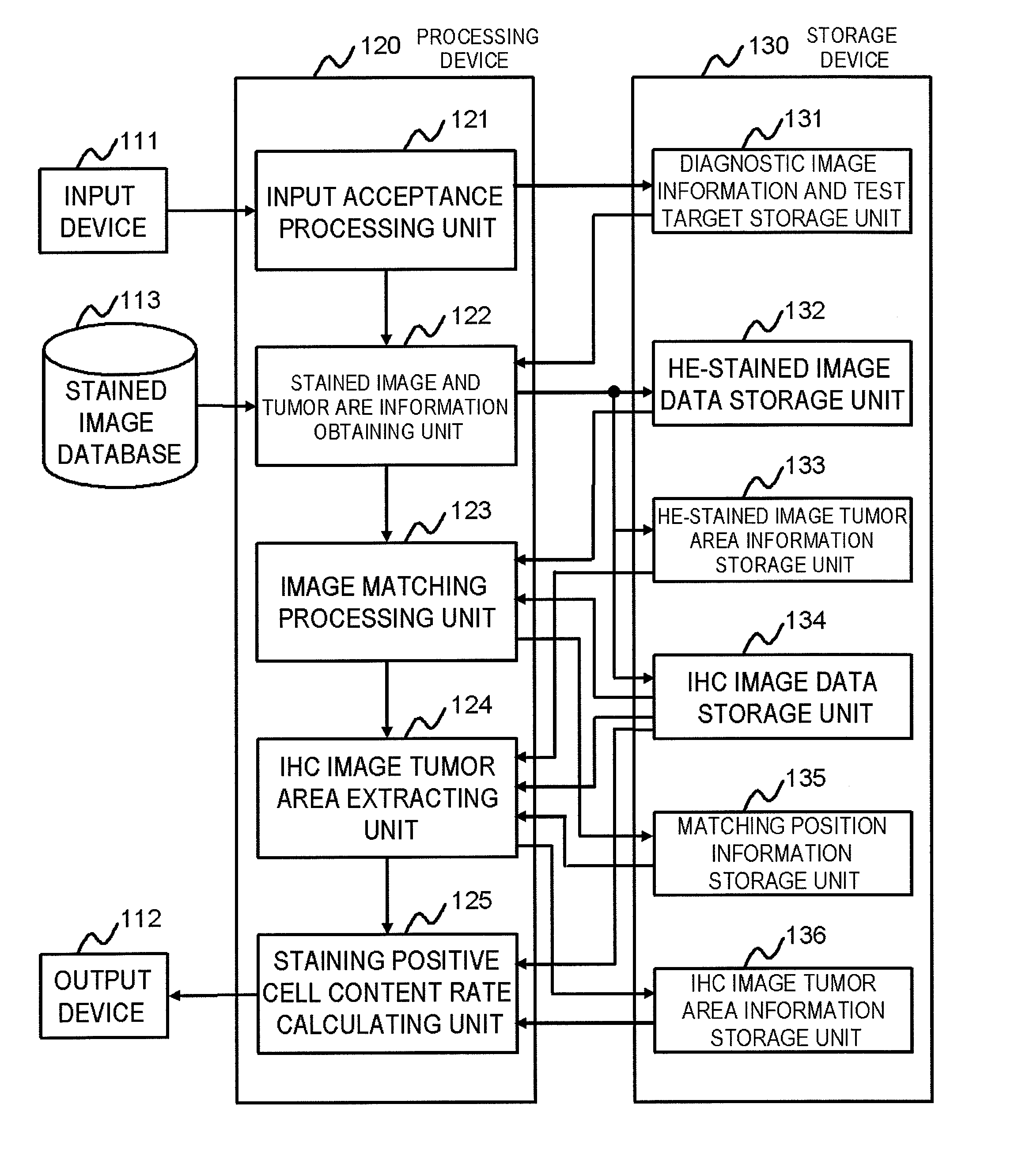
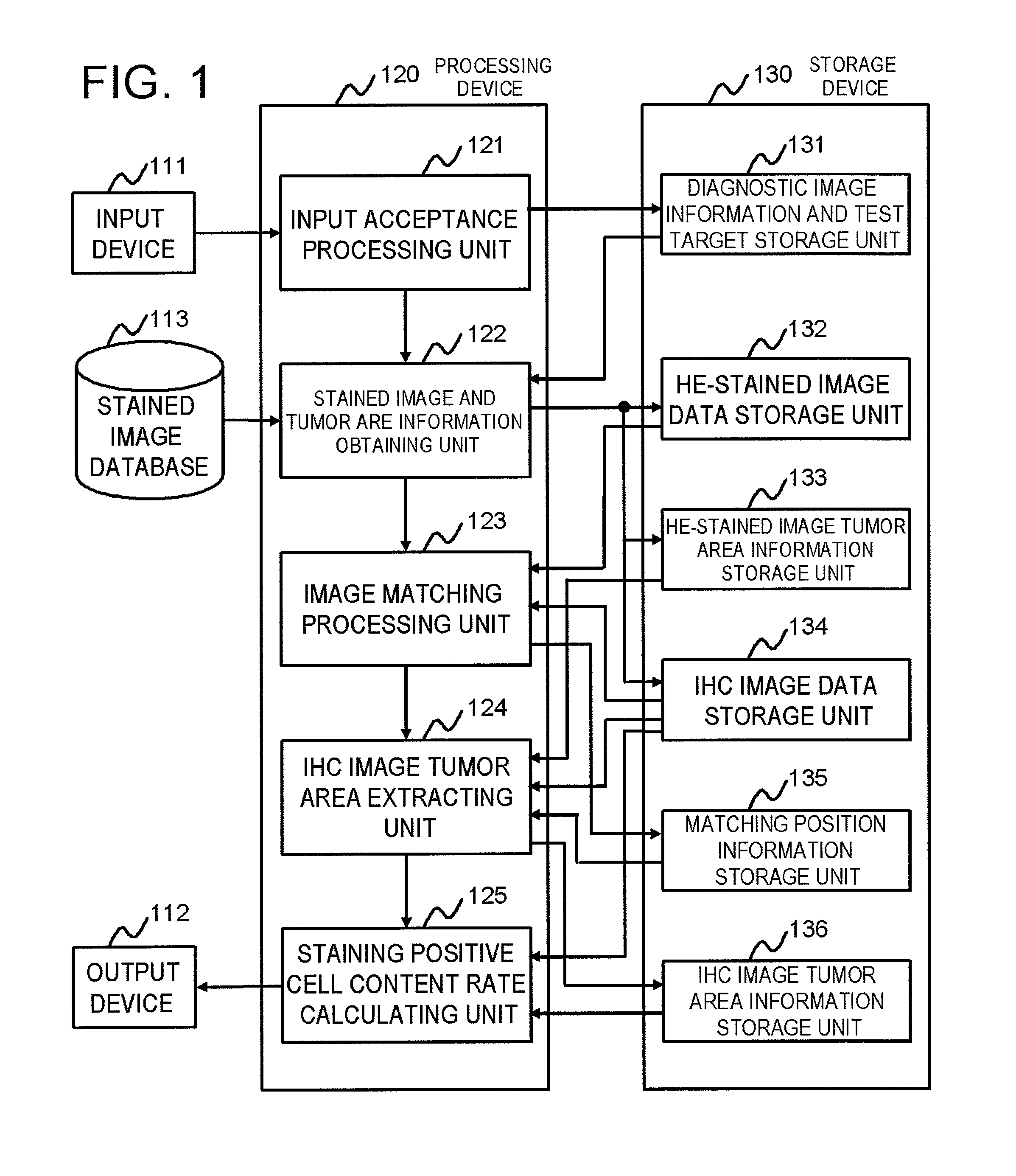
[0032]FIG. 1 is a block diagram showing a first exemplary embodiment of a breast cancer pathological image diagnosis support system according to the present invention. This system is a system for supporting a diagnosis of breast cancer based on a pathological image, and includes an image obtaining unit which obtains an HE-stained image and an IHC image as the pathological images to be diagnosed, an information obtaining unit which obtains information of a tumor area in the HE-stained image, a matching unit which calculates a matching position of the HE-stained image and the IHC image obtained by the image obtaining unit, a specifying unit which specifies a tumor area in the IHC image based on the information of the tumor area in the HE-stained image obtained by the information obtaining unit and information of the matching position calculated by the matching unit, and a calculating unit which calculates a staining positive cell content rate in the tumor area based on information of ...

second exemplary embodiment

[0068]FIG. 7 is a block diagram showing a second exemplary embodiment of the breast cancer pathological image diagnosis support system according to the present invention. The system of this exemplary embodiment is different from the system according to the first exemplary embodiment shown in FIG. 1 in that the staining positive cell content rate calculating unit 125 also calculates staining intensity in addition to the staining positive cell content rate, and other configuration and operation are similar to those of the first exemplary embodiment.

[0069]In FIG. 7, a staining positive cell content rate and staining intensity calculating unit 725 reads the IHC image data and the tumor area from the IHC image data storage unit 134 and the IHC image tumor area information storage unit 136, respectively. Then, the staining positive cell content rate and staining intensity calculating unit 725 counts the number of staining positive cell nuclei and the number of staining negative cell nucle...

third exemplary embodiment

[0077]FIG. 11 is a block diagram showing a third exemplary embodiment of the breast cancer pathological image diagnosis support system according to the present invention. The system of this exemplary embodiment differs from the system according to the first exemplary embodiment shown in FIG. 1 in that the system of this exemplary embodiment is provided with a tumor determining and tumor area calculating unit 1126 (tumor area calculating unit), and other configuration and operation are similar to those of the first exemplary embodiment. In addition, one or more HE-stained image, the IHC image, which is the specimen of the serial section adjacent to the specimen of the HE-stained image, and the specimen adjacent information of the above-described HE-stained image and the above-described IHC image are accumulated in the stained image database 113. In this exemplary embodiment, presence of the tumor area information calculated from the above-described HE-stained image or determined by t...


PUM


Abstract

A breast cancer pathological image diagnosis support system for supporting a diagnosis of breast cancer based on a pathological image is provided. The breast cancer pathological image diagnosis support system includes an image obtaining unit which obtains an HE-stained image and an IHC image as pathological images to be diagnosed; an information obtaining unit which obtains information of a tumor area in the HE-stained image; a matching unit which calculates a matching position of the HE-stained image and the IHC image obtained by the image obtaining unit; a specifying unit which specifies a tumor area in the IHC image based on the information of the tumor area in the HE-stained image obtained by the information obtaining unit and information of the matching position calculated by the matching unit; and a calculating unit which calculates a staining positive cell content rate in the tumor area based on information of the tumor area in the IHC image specified by the specifying unit.

Description

TECHNICAL FIELD[0001]The present invention relates to a system, a method, a program and a recording medium for supporting a diagnosis of breast cancer based on a pathological image.BACKGROUND ART[0002]A pathological diagnosis is performed by viewing a microscope glass specimen referred to as a preparation with a microscope. The microscope specimen, which is made of an organ, tissue and a cell collected from a patient, is obtained by thinly slicing a sample into a few micron thick and thereafter staining the same. A microscopic observation of the specimen supports the diagnosis of illness by a doctor, and is used to determine a future approach to treatment and a prognosis. Especially, an observation of immunohistochemical staining (IHC, also simply referred to as immunostaining) in the breast cancer is used to determine a future indication for endocrine therapy.[0003]An effect indicator of the endocrine therapy in the breast cancer is determined by occupancy or the like of estrogen r...

Claims


Application Information

Patent Timeline
Patent Type & Authority Applications(United States)
IPC IPC(8): G06K9/00
CPCA61B5/055G01N33/57415G01N1/30G06T7/0012
Inventor YAMASHITA, YOSHIKOOKAJIMA, KENJISATO, AKIRA
Owner NEC CORP
Why Eureka
Try Eureka