Chemokines as adjuvants of immune response

a technology of chemokines and adjuvants, applied in the field of human chemokine receptor agonists and antagonists, can solve the problems of complex signals which regulate the trafficking of dendritic cells, are not fully understood, and little information is available regarding the migratory capacity of plasmacytoid dendritic cells

Inactive Publication Date: 2010-04-08
SCHERING CORP
View PDF0 Cites 1 Cited by
  • Summary
  • Abstract
  • Description
  • Claims
  • Application Information

AI Technical Summary

Benefits of technology

[0011]Thus, the invention provides a method of treating disease states comprising administering to an individual in need thereof an amount of a chemokine receptor agonist or antagonist sufficient to increase or decrease the migration of plasmacytoid dendritic cells to the site of antigen delivery.

Problems solved by technology

Signals which regulate the trafficking of dendritic cells, however, are complex and not fully understood.
In particular, very little information is available regarding the migratory capacity of plasmacytoid dendritic cells.

Method used


Image

  • Chemokines as adjuvants of immune response
  • Chemokines as adjuvants of immune response
  • Chemokines as adjuvants of immune response

Examples


example 1

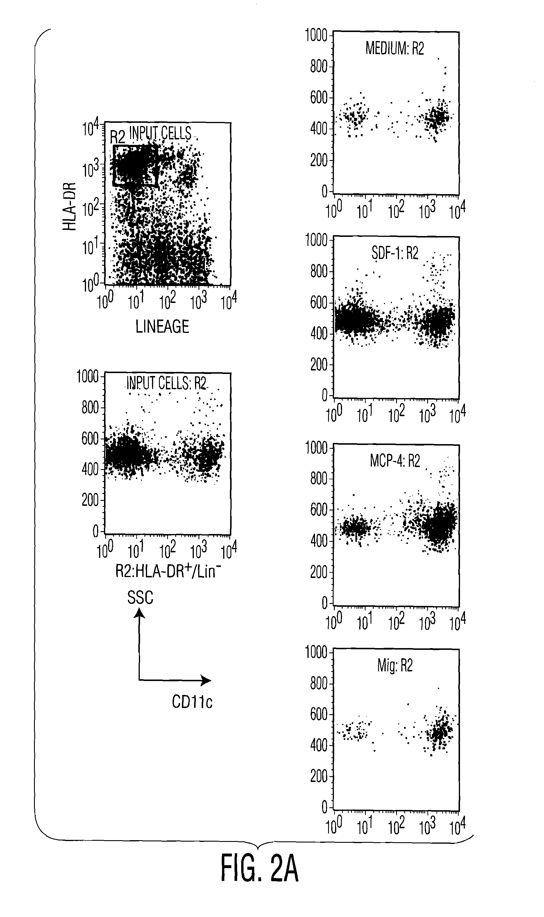
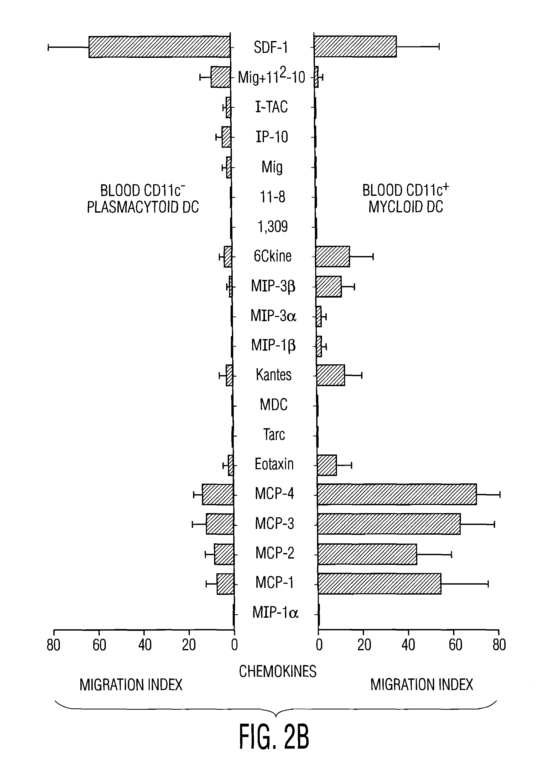
Despite Expression of Receptors for Inflammatory Chemokines, Plasmacytoid DC Respond to the Constitutive Chemokine SDF-1

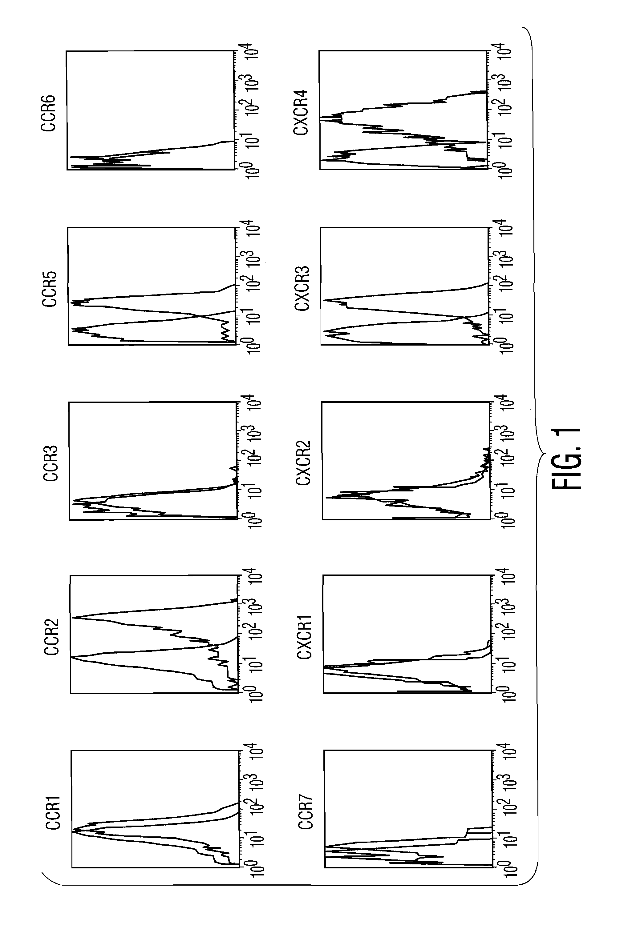
[0105]pDC were enriched from PBMC by magnetic beads depletion. Chemokine receptor and other marker expression was determined by triple staining on enriched blood DC populations and gating on Lin−, CD11c− (FITC), HLA-DR+ (tricolor), using PE-coupled antibodies. Following this protocol, the CD11c− pDC were 95-98% CD45RA+ and IL-3Rα+. pDC expressed CCR2 and CCR5 (FIG. 1) at a comparable levels to CD11c+circulating blood DC (Vanbervliet et al., 2001, Eur J Immunol. 32(1):231-42). CCR1, CCR3, CCR4, CCR6, CCR7, CXCR1, CXCR2, CXCR5 were not significantly expressed as detected by cytofluorimetry (FIG. 1) and / or RT-PCR.

[0106]To determine migration of pDC in response to various chemokines, circulating blood DC subsets were enriched by magnetic bead depletion. After purification, cells were rested for 2 hours at 37° C. and studied in transwell (5 μm pore size) migration assay...

example 2

Plasmacytoid DC Express High Levels of CXCR3 Compared to Other DC Populations

[0110]For blood CD11c− pDC, chemokine receptor and other marker expression was determined by triple staining on enriched blood DC populations and gating on Lin−, CD11c− (FITC), HLA-DR+ (tricolor), using PE-coupled antibodies. Following this protocol the CD11c− pDC were 95-98% CD45RA+ and IL-3Rα+.

[0111]For blood CD11c+ myeloid DC, chemokine receptor and other marker was determined by triple staining gated on Lin−, CD45RA− (FITC), HLA-DR+ (tricolor), using PE-coupled antibodies. Following this protocol the CD11c+myeloid DC were 95-98% CD11c+, IL-3Rα−.

[0112]CD34-derived DC or Monocyte-derived DC were processed for double staining using FITC-conjugated CD1a or CD14 and PE-conjugated monoclonal antibodies against human chemokine receptors.

[0113]As shown in FIG. 1, pDC expressed high levels of CXCR3 at cell surface. In contrast, circulating CD11c+blood DC, as well as other DC populations, did not express signific...

example 3

CXCR3 Ligands Synergize with SDF-1 to Induce Potent Migration of pDC

[0116]Migration assays were performed in response to different SDF-1 and CXCR3 ligand combinations.

[0117]As shows in FIG. 5, in presence of sub-optimal dose of SDF-1 (10 ng / ml) the activity of all 3 CXCR3-ligands was observed at lower concentration (100-500 ng / ml) (FIG. 5B). In addition, when tested in combination with SDF-1, all 3 CXCR3-ligands allowed to lower the threshold of SDF-1 sensitivity by 2 order of magnitude.


PUM

PropertyMeasurementUnit
concentrationsaaaaaaaaaa
timeaaaaaaaaaa
concentrationaaaaaaaaaa

Abstract

Dendritic cells play a critical role in antigen-specific immune responses. Materials and Methods are provided for treating disease states, including cancer, infectious diseases, autoimmune diseases, transplantation, and allergy by facilitating or inhibiting the migration or activation of a specific subset of antigen-presenting dendritic cells known as plasmacytoid dendritic cells (pDC). In particular, methods for treating disease states are provided comprising administration of chemokine receptor agonists and antagonists, alone or in combination with a disease-associated antigen, with or without an activating agent.

Description

FIELD OF THE INVENTION[0001]The invention relates to the use of human chemokine receptor agonists and antagonists in the treatment of disease states, including cancer. The administered chemokine receptor agonists and antagonists direct or prevent the migration of a specific subset of dendritic cells. In one embodiment, disease-specific antigen(s) and / or a moiety designed to activate dendritic cells is administered in conjunction with the chemokine receptor agonist(s).BACKGROUND OF THE INVENTION[0002]Dendritic cells (DC) specialize in the uptake of antigen and their presentation to T cells. DC thus play a critical role in antigen-specific immune responses.[0003]DC are bone marrow-derived and migrate as precursors through bloodstream to tissues, where they become resident cells such as Langerhans cells in the epidermis. In the periphery, following pathogen invasion, immature DC such as Langerhans cells are recruited to the site of inflammation (Kaplan et al., 1992, J. Exp. Med. 175:17...

Claims


Application Information

Patent Timeline
Patent Type & Authority Applications(United States)
IPC IPC(8): A61K39/00C12N5/07A61K38/00A61K38/19A61K39/02A61K39/12A61K39/35A61K39/39A61K45/00A61P31/04A61P31/10A61P31/12A61P33/00A61P35/00A61P37/00A61P37/06A61P37/08A61P43/00C12N5/077C12N5/0783C12N5/0784C12N5/09
CPCA61K39/39A61K2039/55511A61K2039/55522A61K38/202A61K38/191A61K38/195A61K38/212A61K2300/00A61P31/04A61P31/10A61P31/12A61P33/00A61P35/00A61P37/00A61P37/06A61P37/08A61P43/00
Inventor CAUX, CHRISTOPHEVANBERVLIET, BEATRICEPATUREL, CARINEVICARI, ALAINTRINCHIERI, GIORGIOBRIERE, FRANCINEBENDRISS, NATHALIE
Owner SCHERING CORP
Why Eureka
Try Eureka