
Method and apparatus for localizing an ultrasound catheter

a catheter and ultrasound technology, applied in the field of medical imaging systems, can solve problems such as heart arrhythmias, problems such as the difficulty of adjusting the catheter,

Active Publication Date: 2010-04-29
ST JUDE MEDICAL ATRIAL FIBRILLATION DIV
View PDF79 Cites 98 Cited by
  • Summary
  • Abstract
  • Description
  • Claims
  • Application Information

AI Technical Summary

Benefits of technology

The present invention provides an imaging system with an ultrasound catheter that can generate positional information and has a restricted field of view. The system can also coregister images from the catheter with positional information and display them based on the calculated position. Additionally, the invention provides a method for generating and displaying medical images from a catheter-based imaging ultrasound sensor with a restricted field of view. The technical effects of the invention include improved accuracy and efficiency in medical imaging and reduced risk of complications during procedures.

Problems solved by technology

The Dupree system, however, may be problematic in some applications due to its use of an electrode generated electric field to determine the location of the probe.
In particular, electric fields generated intra-body can generate electrical currents which flow in the body that may cause muscle stimulation, which may result in heart arrhythmias, etc., when used in or near the heart, such as intra-cardiac sensing or treatment.

Method used


Image

  • Method and apparatus for localizing an ultrasound catheter
  • Method and apparatus for localizing an ultrasound catheter
  • Method and apparatus for localizing an ultrasound catheter

Examples


Embodiment Construction

[0020]Reference will now be made in detail to exemplary embodiments of the present invention. Wherever possible, the same reference numbers will be used throughout the drawings to refer to the same or like parts.

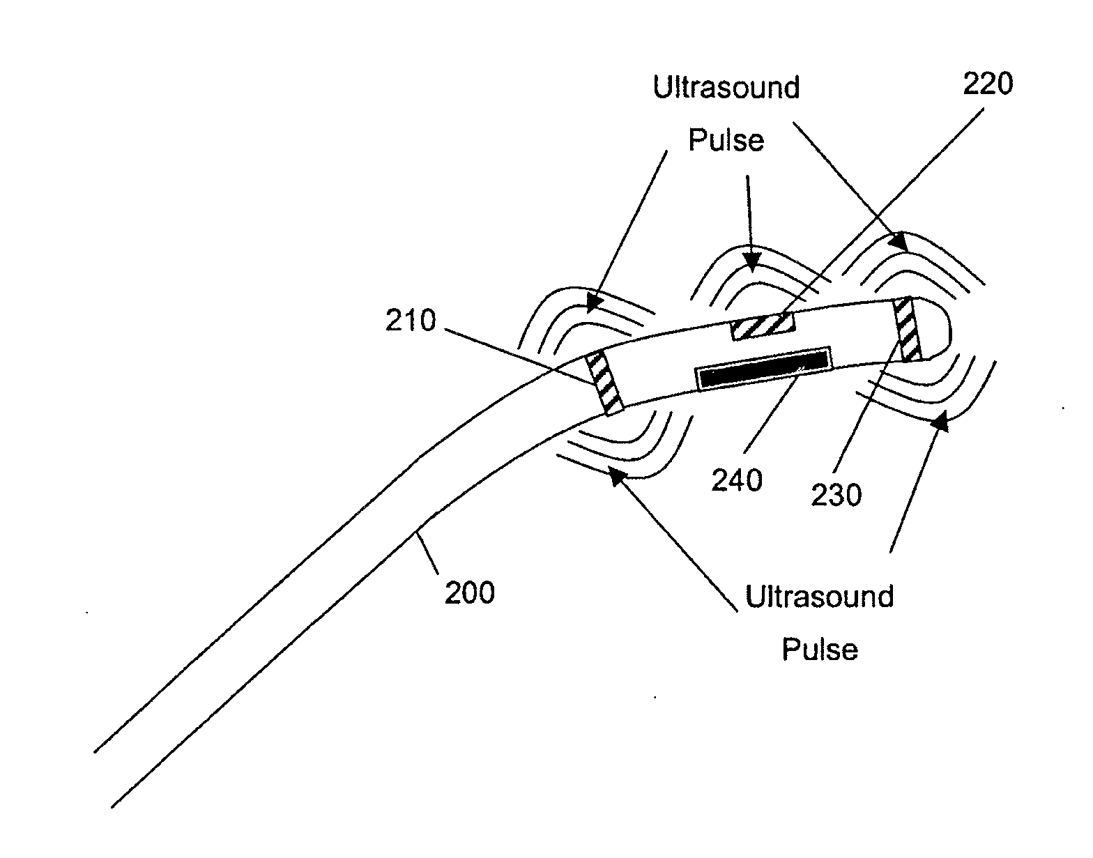
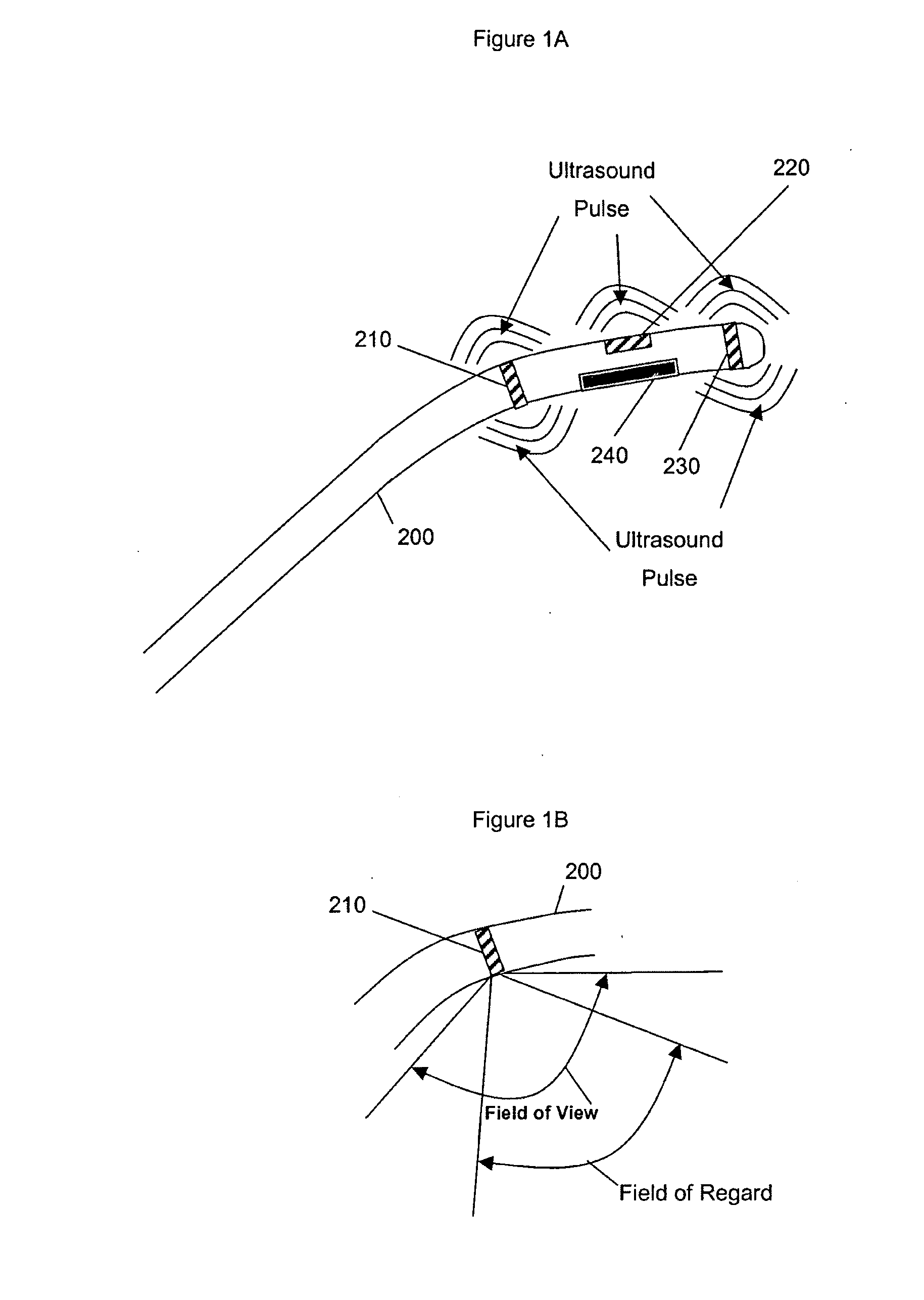
[0021]The various embodiments of the present invention provide capabilities to determine the location of medical instrumentation and / or treatment devices within a patient's body using ultrasound echolocation and / or three dimensional (3D) triangulation techniques, and to use this localized information in conjunction with medical images. Relative positions of one instrument with respect to other instruments and registration of instrumentation positions with respect to the patient's body may be obtained, which is generally referred to herein as “localizing” the instrumentation. Thus, references to catheters as particular types of medical instrumentation and treatment devices are not intended to be limiting since the claimed systems and methods equally apply to other non-cathete...


PUM


Abstract

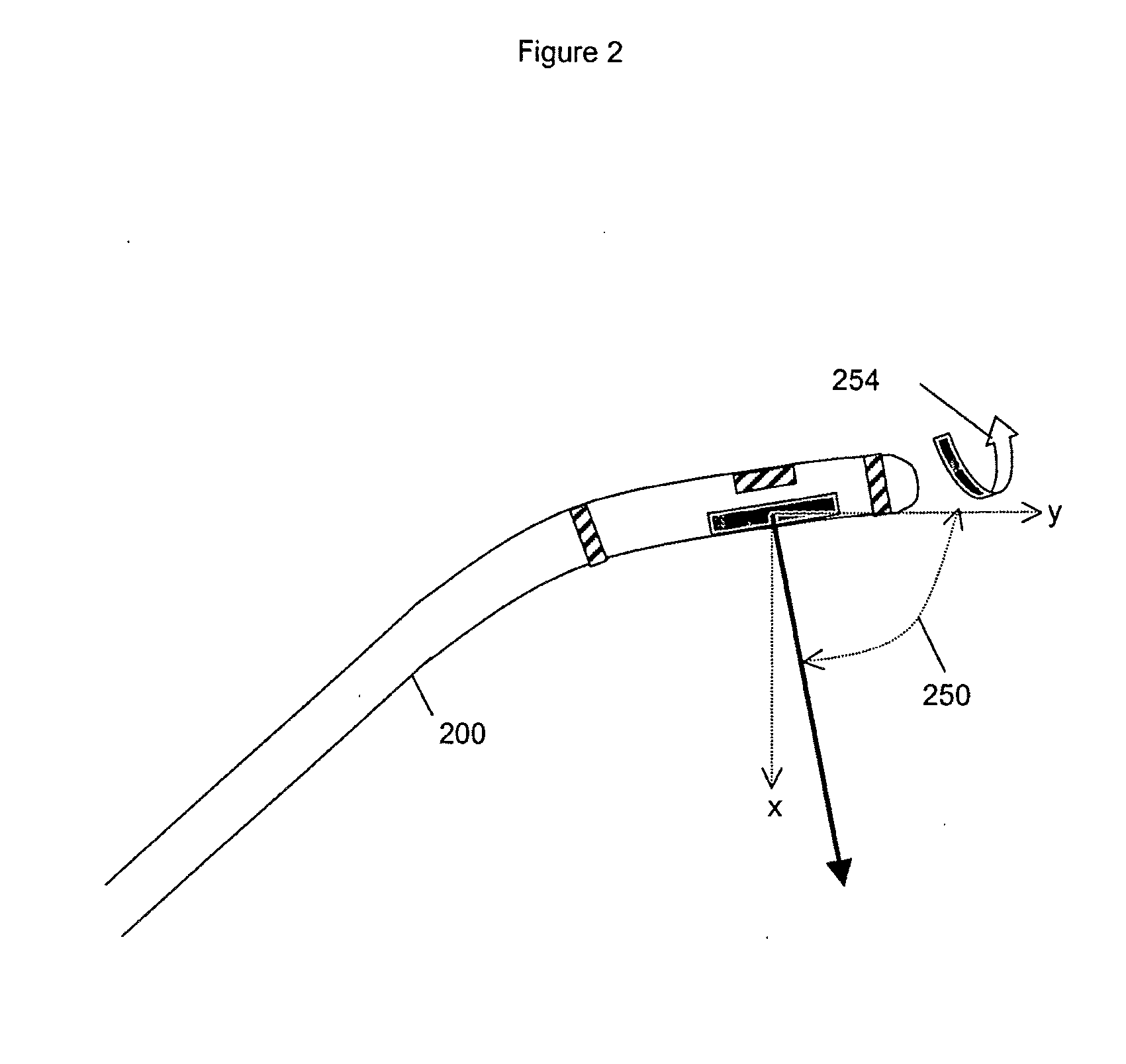
An imaging system is provided with an ultrasound catheter and a controller coupled to the ultrasound catheter. The catheter includes a localizer sensor configured to generate positional information for the ultrasound catheter, and an imaging ultrasound sensor having a restricted field of view. The controller co-registers images from the imaging ultrasound sensor with positional information from the localizer sensor.

Description

CROSS REFERENCE[0001]The present invention is a continuation of U.S. patent application Ser. No. 10 / 994,424 filed 23 Nov. 2004, which is incorporated by reference herein in its entirety.BACKGROUND OF THE INVENTION[0002]1. Field of the Invention[0003]The present invention relates generally to medical imaging systems, and more particularly to a method and apparatus for localizing an ultrasound imaging catheter.[0004]2. Description of the Related Art[0005]Medical imaging technology is used to improve the diagnosis and treatment of medical conditions. Presently available medical imaging technology includes a wide variety of ultrasound, X-ray, nuclear, magnetic resonance imaging (MRI) and other imaging systems. A technology of particular benefit to diagnosis and treatment of cardiovascular conditions uses imaging ultrasound detectors mounted a percutaneous catheter.[0006]Techniques exist for localizing catheters deployed within a patient's body. One such technique is described in U.S. Pa...

Claims


Application Information

Patent Timeline
Patent Type & Authority Applications(United States)
IPC IPC(8): A61B5/05A61B8/14
CPCA61B5/06A61B5/064A61B8/12A61B8/5238A61B8/463A61B5/063A61B8/445A61B8/4263A61B8/4254A61B8/54A61B5/062A61B8/4488
Inventor BYRD, CHARLES BRYANDALA-KRISHNA, PRAVEENJENKINS, DAVID A.
Owner ST JUDE MEDICAL ATRIAL FIBRILLATION DIV
Learn More