Method and System for Left Ventricle Detection in 2D Magnetic Resonance Images Using Ranking Based Multi-Detector Aggregation

a technology of multi-detector aggregation and ranking, applied in the field of medical imaging of the heart, can solve the problems of large flexibility, difficult automatic lv detection, and difficult automatic lv detection, and achieve the effect of avoiding the lv long axis orientation constraint, and avoiding the lv long axis

Inactive Publication Date: 2010-06-10
SIEMENS HEATHCARE GMBH
View PDF16 Cites 15 Cited by
  • Summary
  • Abstract
  • Description
  • Claims
  • Application Information

AI Technical Summary

Problems solved by technology

Automatic LV detection in MRI images is a challenging problem due to large variations in orientation, size, shape, and image intensity of the LV.
However, this flexibility presents a large challenge for automatic LV detection because both the position and orientation of the LV are unconstrain

Method used


Image

  • Method and System for Left Ventricle Detection in 2D Magnetic Resonance Images Using Ranking Based Multi-Detector Aggregation
  • Method and System for Left Ventricle Detection in 2D Magnetic Resonance Images Using Ranking Based Multi-Detector Aggregation
  • Method and System for Left Ventricle Detection in 2D Magnetic Resonance Images Using Ranking Based Multi-Detector Aggregation

Examples


Embodiment Construction

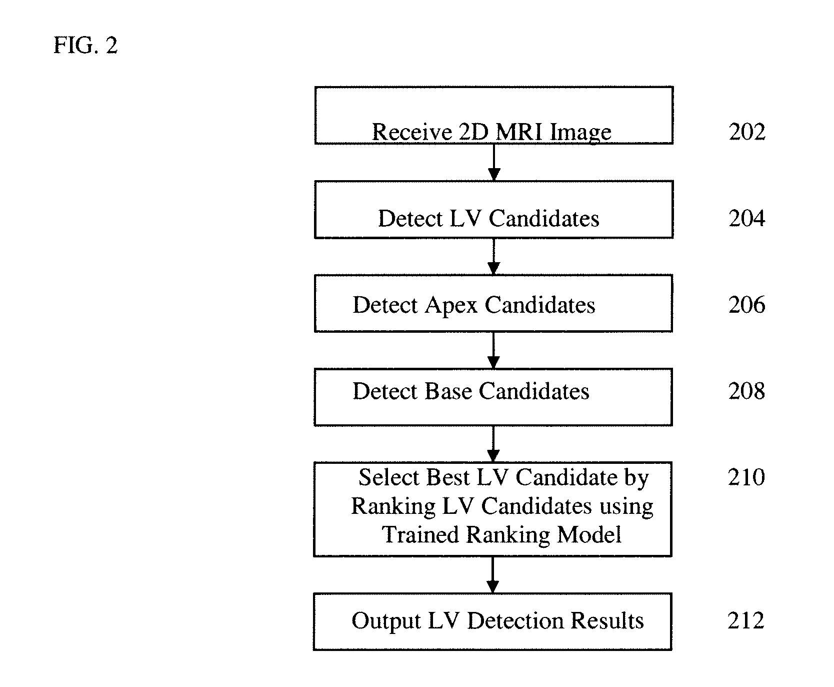
[0014]The present invention is directed to a method and system for automatic left ventricle (LV) detection in 2D magnetic resonance imaging (MRI) images. Embodiments of the present invention are described herein to give a visual understanding of the left ventricle detection method. A digital image is often composed of digital representations of one or more objects (or shapes). The digital representation of an object is often described herein in terms of identifying and manipulating the objects. Such manipulations are virtual manipulations accomplished in the memory or other circuitry / hardware of a computer system. Accordingly, it is to be understood that embodiments of the present invention may be performed within a computer system using data stored within the computer system.

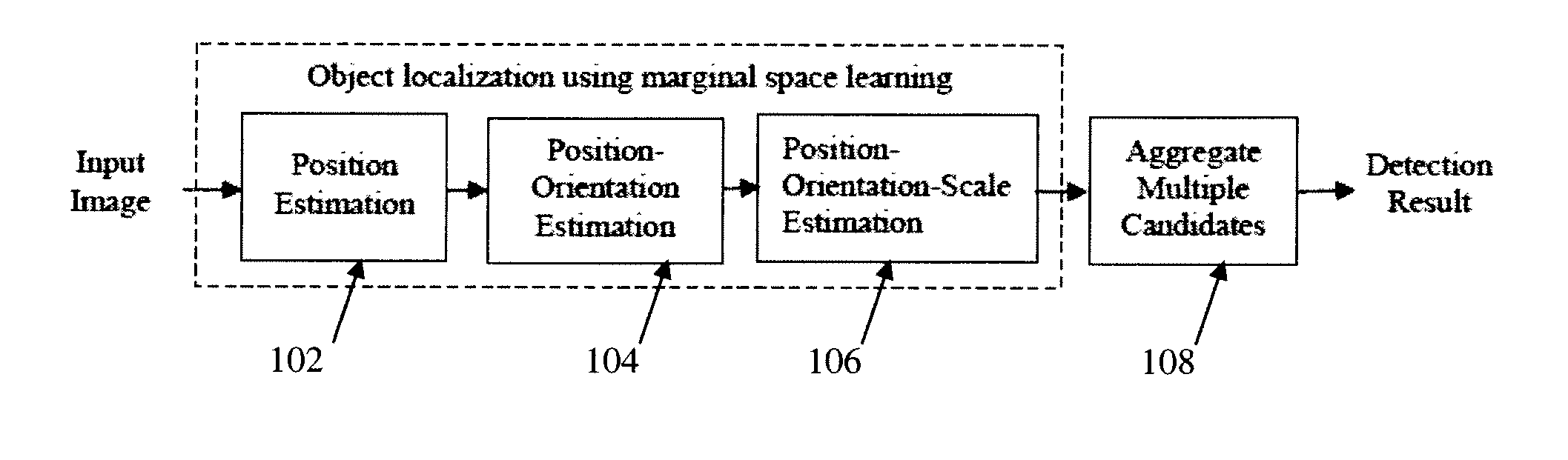
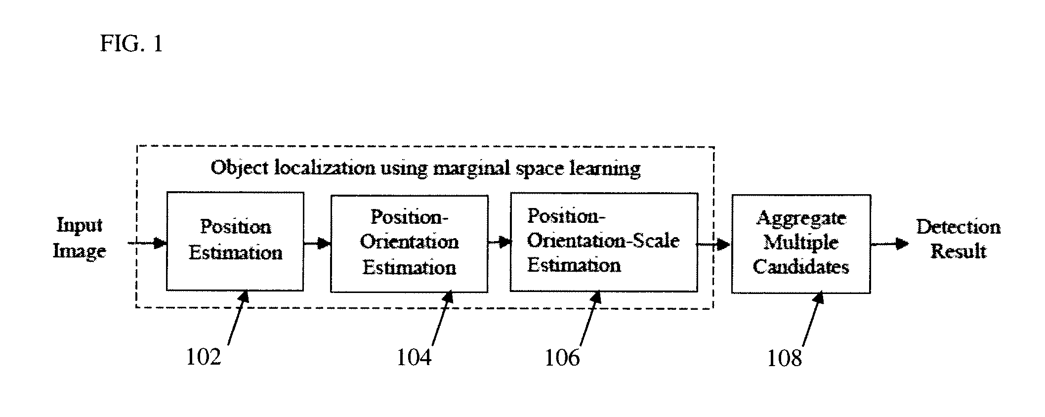
[0015]Discriminative learning based approaches are efficient and robust for solving many 2D detection problems. In such methods, shape detection and localization is formulated as a classification problem: wheth...


PUM


Abstract

A method and system for left ventricle (LV) detection in 2D magnetic resonance imaging (MRI) images is disclosed. In order to detect the LV in a 2D MRI image, a plurality of LV candidates are detected, for example using marginal space learning (MSL) based detection. Candidates for distinctive anatomic landmarks associated with the LV are then detected in the 2D MRI image. In particular, apex candidates and base candidates are detected in the 2D MRI image. One of the LV candidates is selected as a final LV detection result by ranking the LV candidates based on the LV candidates, the apex candidates, and the base candidates using a trained ranking model.

Description

[0001]This application claims the benefit of U.S. Provisional Application No. 61 / 120,143, filed Dec. 5, 2008, the disclosure of which is herein incorporated by reference.BACKGROUND OF THE INVENTION[0002]The present invention relates to medical imaging of the heart, and more particularly, to automatic detection of the left ventricle in 2D magnetic resonance images.[0003]Cardiovascular disease is the leading cause of death in developed countries. Early diagnosis can be effective in reducing the mortality of cardiovascular disease. Magnetic resonance imaging (MRI) can accurately depict cardiac structure, function, perfusion, and myocardial viability with a capacity unmatched by any other imaging modality. Accordingly, MRI is widely accepted as the gold standard for heart chamber quantification, which means that measurements extracted using other imaging modalities, such as echocardiography and computed tomography (CT), typically must be verified using MRI. Quantification of the left ve...

Claims


Application Information

Patent Timeline
IPC IPC(8): G06K9/62
CPCG06K9/3241G06K2209/051G06T7/0081G06T2207/30048G06T2207/10088G06T2207/20081G06T7/0087G06T7/11G06T7/143G06V10/255G06V2201/031
Inventor ZHENG, YEFENGGEORGESCU, BOGDANLITTMANN, ARNEMUELLER, EDGARCOMANICIU, DORIN
Owner SIEMENS HEATHCARE GMBH
Why Eureka
Try Eureka