

Method and device for microdialysis sampling
a microdialysis and microdialysis technology, applied in the field of microdialysis sampling, can solve the problem that the design is not intended for organs
Active Publication Date: 2010-06-17
MD BIOMEDICAL
View PDF9 Cites 4 Cited by
- Summary
- Abstract
- Description
- Claims
- Application Information
AI Technical Summary
Problems solved by technology
This design has a clear disadvantage compared with the present invention because it does not incorporate a method that allows for microdialysis sampling by the placem
Method used
View moreImage
Smart ImageViewing Examples

Examples
 PUM
 Abstract
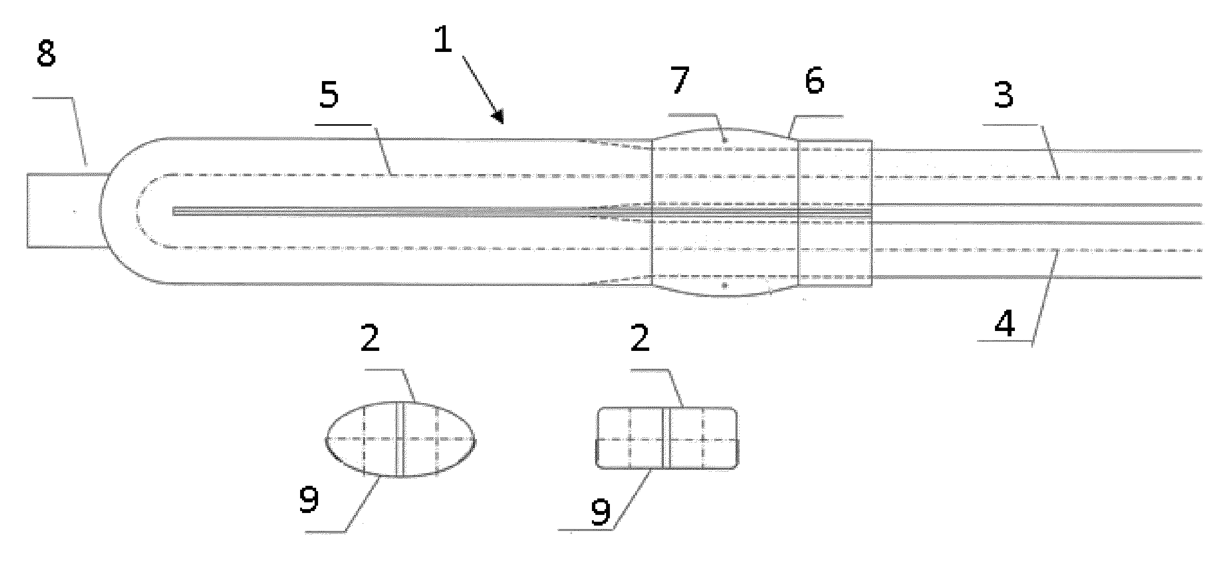
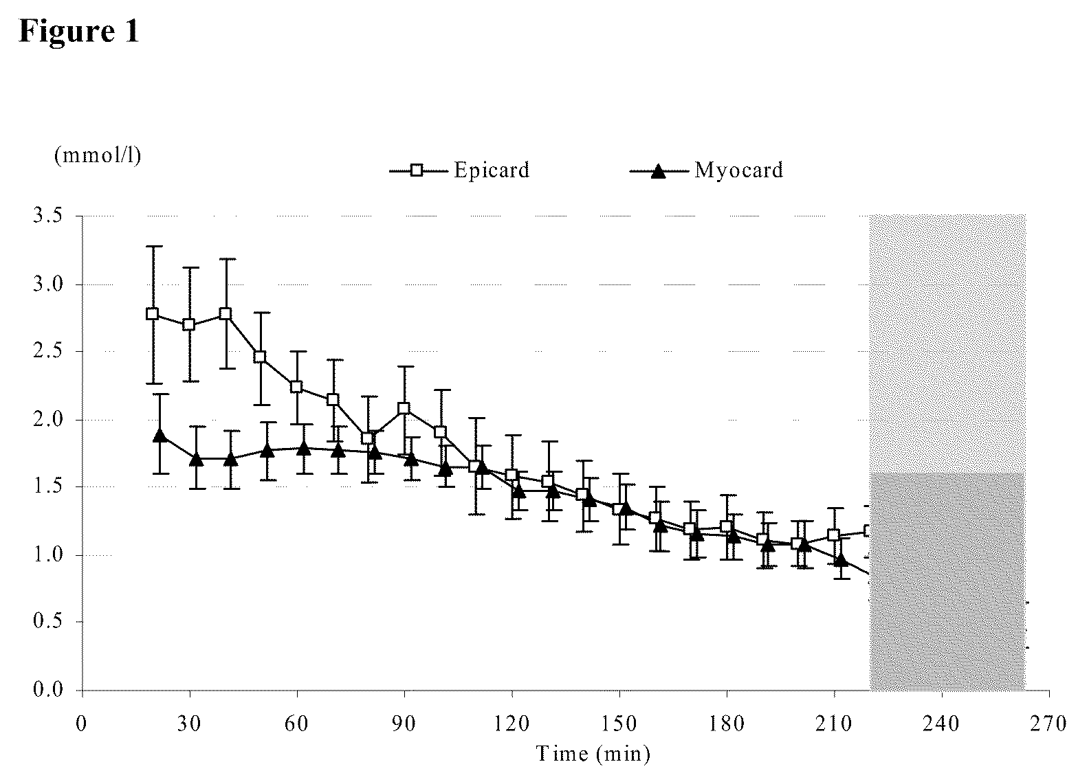
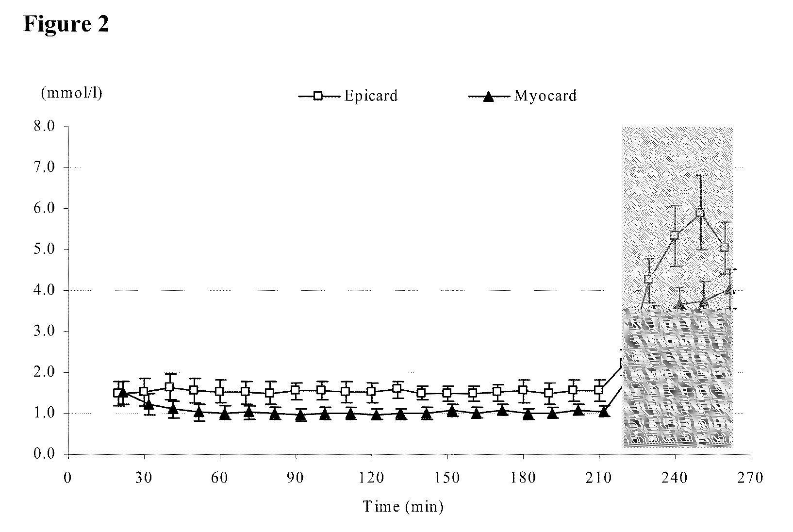
The microdialysis technique and device of the present invention can be used to study metabolic aspects of human and animal organs. When a microdialysis catheter is for example placed into the substance of a beating heart, there is always concern for problems with the catheter position. Further, there is always a risk for damage to heart tissue as well as to the catheter. The present invention is designed to avoid such damages, and a study has confirmed that data obtained from the epicardial probe of the present invention reflects the state of the myocardial metabolism. In anesthetized normoventilated pigs (n=20), a sternotomy was performed and a Gore-tex suture snare was placed around a branch of the left anterior descending (LAD) artery. One microdialysis probe was inserted in the myocardial tissue supplied by the snared LAD branch. Another microdialysis probe was placed on the epicardial surface above the myocardial probe. The protocol included four, ten minute ischemic periods (intervention group) followed by a forty minute ischemic period. The control group was only exposed to a forty minute ischemic period. It was possible to place the microdialysis probes of the present invention on the epicardial surface, and to recover samples reflecting the myocardial metabolic state of the heart. It was also possible to measure rapid changes in concentrations especially for lactate, where ischemia periods as short as ten minutes resulted in detectable increases. A similar pattern was found when analysing glucose and glycerol concentrations in paired samples. Data obtained from the device and method of the present invention, placed on the epicardial surface showed a similar pattern compared to data from a standard probe placed in the myocardium. Thus, the epicedial application of the present invention is useful for perioperative monitoring during cardiac surgery.
Description
TECHNICAL FIELD[0001]The present invention concerns a method and device for microdialysis sampling the well-being or metabolic conditions of vital organs within humans and animals. More specifically the present invention regards the measurement of substance concentrations (molecules) on the surface of vital organs in accordance with the claims.BACKGROUND OF THE INVENTION[0002]Microdialysis is a means of sampling substances from the body to help clinicians assess well-being or metabolic conditions by providing serial biochemical samples from a catheter which lies within the substance of an organ. Current commercial microdialysis systems are equipped for sample collection, handling, and analysis of small molecules including glucose, lactate, pyruvate and glycerol as markers of cell injury. Sample collection is based on passive diffusion through a semi-permeable membrane placed at the end of a catheter.[0003]Microdialysis sampling can be performed on organs which move. For example micr...
Claims
Login to View More Application Information
Patent Timeline
 IPC IPC(8): A61B5/00
CPCA61B5/14525A61B5/14528A61B10/0045A61M2025/0286A61M25/00A61M25/02A61B2010/008
InventorABRAHAMSSON, PERNILLA BIRGITTA
OwnerMD BIOMEDICAL