
Phantom for ultrasound guided needle insertion and method for making the phantom

Inactive Publication Date: 2010-08-05
KONINKLIJKE PHILIPS ELECTRONICS NV
View PDF9 Cites 46 Cited by
  • Summary
  • Abstract
  • Description
  • Claims
  • Application Information

AI Technical Summary

Benefits of technology

[0013]Thanks to the invention, the phantom permits a realistic simulation of the human body behavior during an ultrasound guided needle insertion in a blood vessel, since the phantom comprises a particular material for mimicking each particular part of the human body site, the materials being arranged to reproduce the mechanical as well as the ultrasound properties of the corresponding (mimicked) parts of an actual human body. Therefore, the phantom mechanically behaves as a human body and permits a realistic ultrasound imaging of the needle insertion. In other words, the phantom of the invention enables to simulate the anatomy of a specific human body site, the deformation behavior of the blood vessels and their surroundings and the ultrasound properties of the blood vessels, tissues and skin when inserting a needle into a blood vessel. The phantom is adapted for simulating a manual as well as an automated needle insertion method.

Problems solved by technology

However, it does not permit to mimic the behavior of a human body site when a needle is inserted in a blood vessel.

Method used


Image

  • Phantom for ultrasound guided needle insertion and method for making the phantom
  • Phantom for ultrasound guided needle insertion and method for making the phantom

Examples


Embodiment Construction

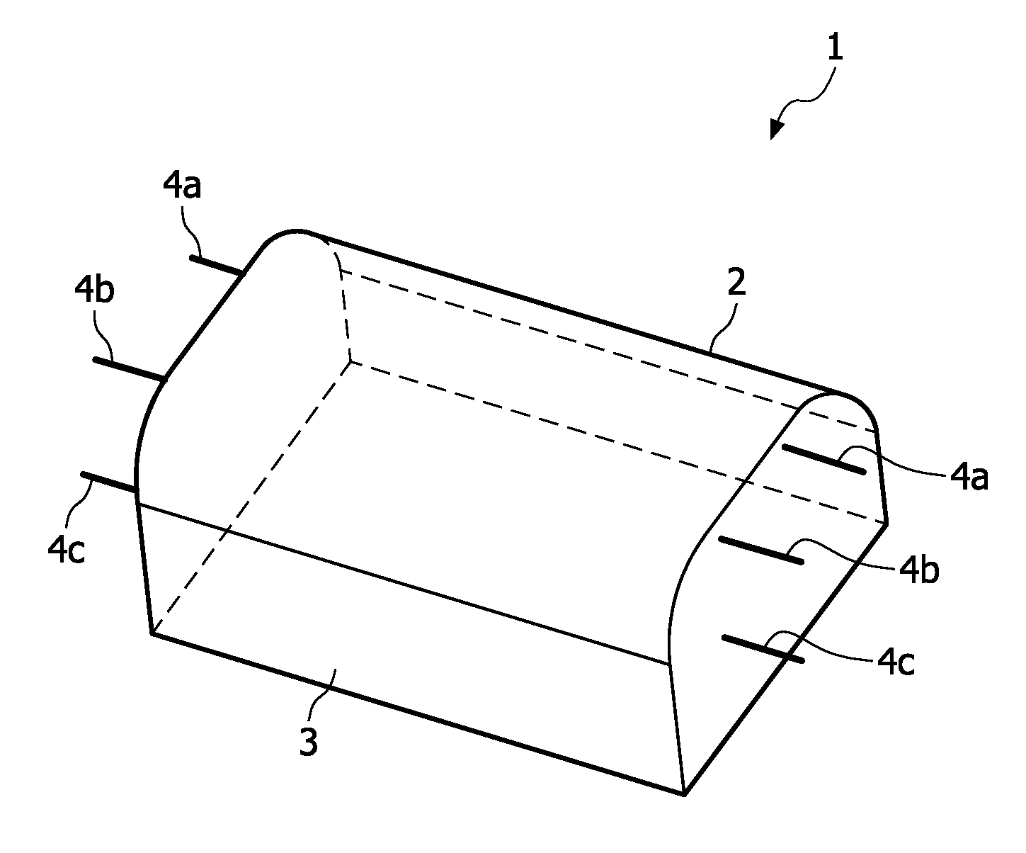
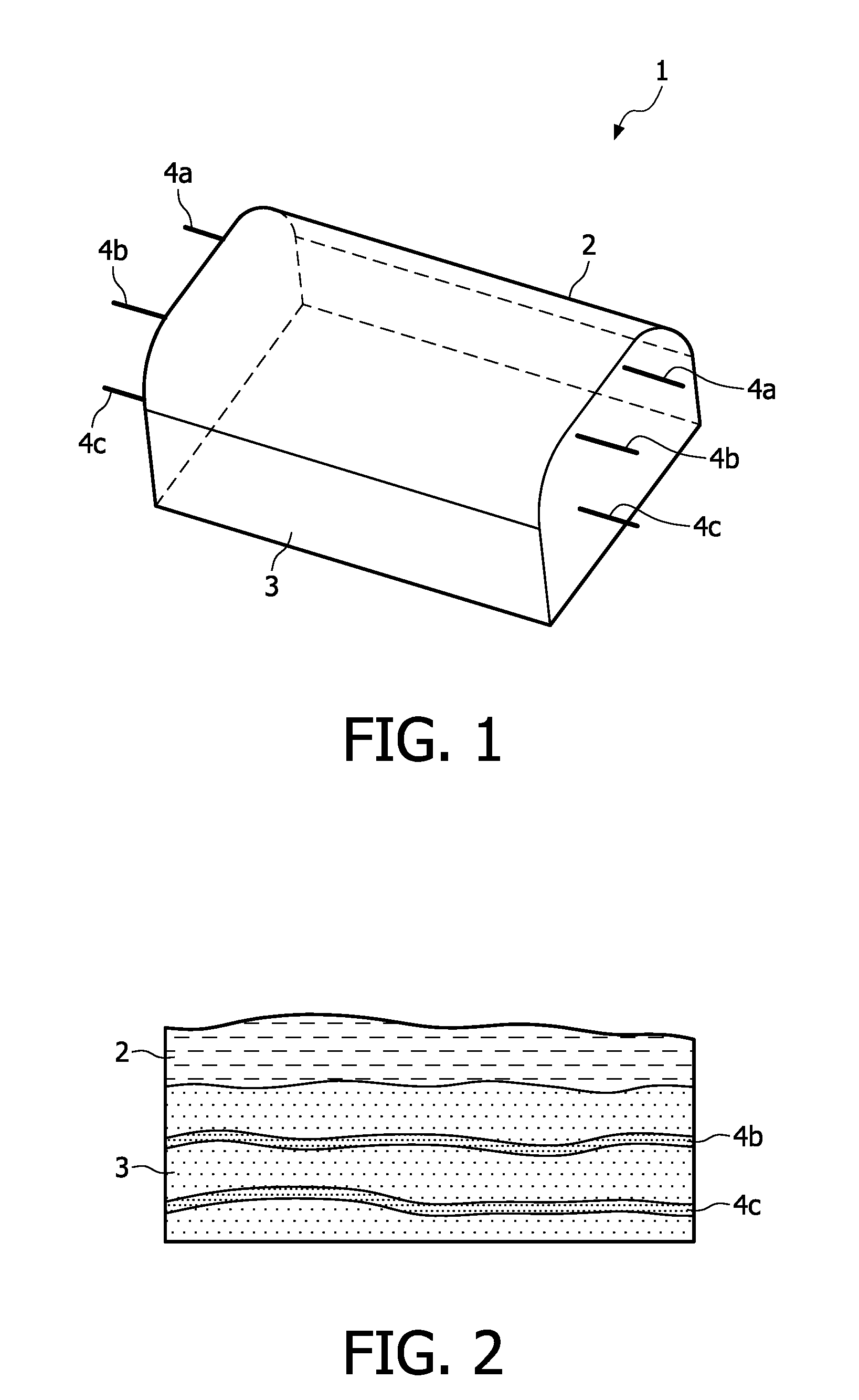
[0030]With reference to FIGS. 1 and 2, a phantom 1 according to the invention comprises a skin mimicking layer 2, a tissue mimicking layer 3 and artificial blood vessels 4a, 4b, 4c. The phantom 1 is a test object that is used in simulation of ultrasound image-guided medical invasive procedures, namely insertion of a needle in a blood vessel of a human body site. In the embodiment described, the phantom 1 mimics the elbow inner region of a human body with its superficial veins, where venipuncture is usually performed. The invention in particular applies to venipuncture, but it more generally applies to any insertion of a needle into a blood vessel of a human body site.

[0031]The skin mimicking layer 2 is formed in a first material, which in this embodiment is latex, in particular fluid latex; the thickness of the skin mimicking layer 2 is substantially equal to the one of skin in the elbow region of a human body. The tissue mimicking layer 3 here mimics a fat layer and is formed in a ...


PUM


Abstract

The invention relates to a phantom for simulating the ultrasound guided insertion of a needle in a blood vessel of a human body site. The phantom comprises: a skin mimicking layer (2), formed in a first material; —a tissue mimicking layer (3), formed in a second material and at least one artificial blood vessel (4a, 4b, 4c), formed in a third material, the first, second and third material being arranged to reproduce both the mechanical and the ultrasound properties of the corresponding parts of the human body site. Thanks to the invention, the phantom permits a realistic simulation of a human body site behavior.

Description

FIELD OF THE INVENTION[0001]The invention relates to needle insertion and more particularly to ultrasound guided insertion.BACKGROUND OF THE INVENTION[0002]Insertion of a needle into a blood vessel of a patient is a very common medical procedure in order, for instance, to gather blood from the vessel or to inject a product such as a vaccine. The insertion of the needle may not always be performed perfectly and monitoring of the insertion may be beneficial. Consequently, there was a great need for ultrasound guided needle insertion.[0003]Ultrasound guided needle insertion may be performed manually or automatically. In the case of manual insertion, a person may hold ultrasound imaging means—such as an ultrasound probe—in one hand and a syringe holding the needle in the other hand; as the needle is inserted, the person can check the movements of the needle in the tissues of the patient on images obtained in real-time thanks to the imaging means. In the case of automated insertion, a de...

Claims


Application Information

Patent Timeline
IPC IPC(8): G09B23/30
CPCG09B23/28
Inventor GEERLIGS, MARIONNEERKEN, SIEGLINDEBEZEMER, ROBERT ALFREDHEKKENBERG, ROBERTUS
Owner KONINKLIJKE PHILIPS ELECTRONICS NV
Learn More