Use of non-clonal chromosomal aberrations for cancer research and clinical diagnosis

a cancer and clinical diagnosis technology, applied in the field of diagnostic or evaluation methods, can solve the problems of incongruity of current cancer concept, difficult data interpretation, complex karyotypical changes that occur, etc., and achieve the effects of stimulating transcriptional activation, promoting proliferation and survival, and high diversity

Inactive Publication Date: 2010-09-16
HENG HENRY H +1
View PDF0 Cites 7 Cited by
  • Summary
  • Abstract
  • Description
  • Claims
  • Application Information

AI Technical Summary

Benefits of technology

[0038]The chromosome represents not only the vehicle in which genes are carried but also serves as a genetic framework that controls all genes in a systemic manner. The karyotypic-defined genome is the driving force for cancer evolution and for other types of organismal evolution. Clinical cytogenetic analysis is commonly done in many types of cancers and often serves as a diagnostic and prognostic marker of cancer progression. Clonal events are recorded, but recent evidence shows that nonclonal events provide the diversity that is required for tumor progression and survival in the extremely harsh environment of a tumor. Change on the chromosomal level has the capability of directly altering the regulation of hundreds or possibly thousands of genes. Chromosomal change, including translocations, deletions, duplications, inversions, defective mitotic figures, and fragmentation of chromosomes can serve to increase diversity that is required for cancer progression and evolution.
[0046]Third, the rate of NCCAs can be easily accessed from patients by screening lymphocytes derived from drawn blood samples rather than direct tissue biopsies. This makes the use of NCCAs a practical and feasible tool. The use of lymphocytes for the purpose of monitoring genetic susceptibility is not new. Lymphocytes have been used to detect chromosomal breakage as a means to correlate increased chromosomal instability in cancer patients. However, the rates of breakage are less reliable than rates of non-clonal translocations. Chromosomal breakage used to predict the likelihood of cancer was successful based on group data, but was not as reliable when used for individual based data. Levels of NCCAs can be used to monitor and establish a genomic instability baseline for each individual (since the genetic background is the same for all of an individual's cells). The use of a system to monitor genomic instability based on the use of NCCAs obtained from patient's lymphocytes is strongly supported by data.
[0049]Studies on clonal diversity and subsequent clinical outcomes in Barrett's esophagus reinforce the concept that cancer progression occurs through somatic evolution driven by genome instability coupled with an increase in or accumulation of clonal diversity. To date, however, most evolutionary analyses have focused on specific genetic loci rather than the overall genome level diversity. The impact of genetic variation at the genome level is much more profound than at the gene level, as the higher level of organization often constrains lower levels and displays more stable characteristics than lower levels. It is therefore expected that the major form of cellular population diversity is generated by karyotypic heterogeneity reflected as NCCA / CCA cycles (previously described as the waves of clonal expansion with the regeneration of genetic diversity in between) occurring during somatic evolution. It is thus more reliable and easier to measure the degree of diversity at the genome level than at the individual gene level. In addition, it has been a challenge to trace individual genes for most cancer types where there is a high level of genomic heterogeneity.
[0083]To exclude the possibility that ploidy rather than a high degree of diversity contribute to the tumorigenicity that is observed with CSC-MCF10A3, an additional subline was selected with identical karyotypes (and ploidy status) but these lines displayed a diversity of NCCAs. This subline was obtained by spontaneously transforming MCF10 cells by over-expression of HOXA 1. Human growth hormone-regulated HOXA 1 has been shown to be a mammary epithelial oncogene. HOXA 1 stimulates the transcriptional activation of a number of pro-oncogenic molecules including cyclin D1 and Bcl-2 that promotes proliferation and survival. Over-expressed HOXA 1 in human mammary carcinoma cells results in drastically increased tumorigenicity.

Problems solved by technology

The challenge for this approach is the complexity of the karyotypical changes that occur and the karyotypical heterogeneity that is associated with cancer cell lines and tumor samples.
Similarly, the heterogeneity of tumor samples makes data interpretation difficult with significant exceptions occurring when various samples of the same tumors were analyzed.
The technical challenge for genome structure and function studies by using this approach is to develop a system that can monitor the genome structure and changes caused by these targeted genes.
Further, the list of cancer genes continues to grow, which brings in to question the goals and rationale of continuing to attempt to identify a handful of commonly shared gene mutations in cancer.
Clearly, the current concept of cancer is not consistent with the reality of the presence of high degrees of genetic diversity in patients.
Unfortunately, this highly anticipated approach is delivering unwanted results.

Method used


Image

  • Use of non-clonal chromosomal aberrations for cancer research and clinical diagnosis
  • Use of non-clonal chromosomal aberrations for cancer research and clinical diagnosis
  • Use of non-clonal chromosomal aberrations for cancer research and clinical diagnosis

Examples


Embodiment Construction

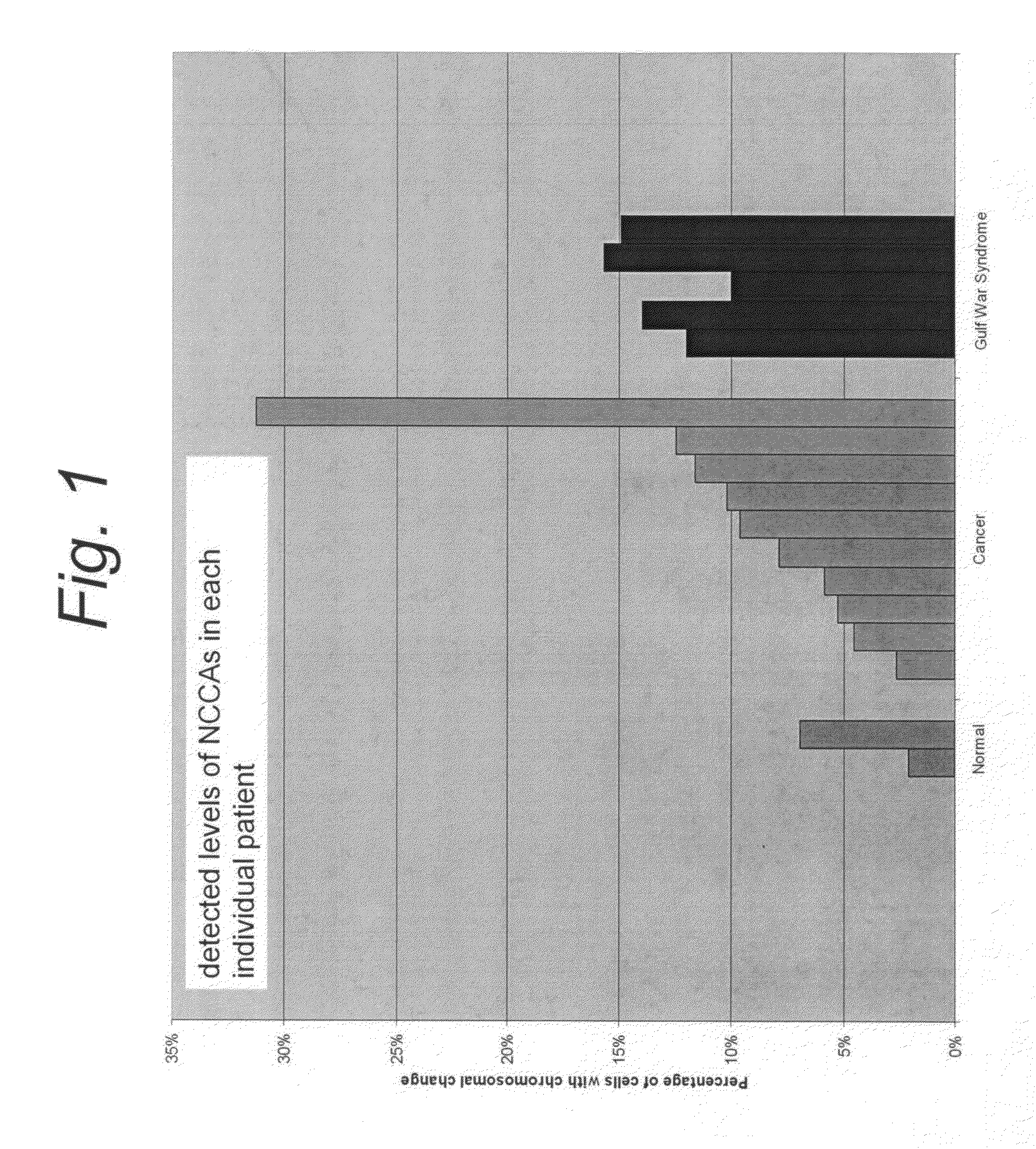
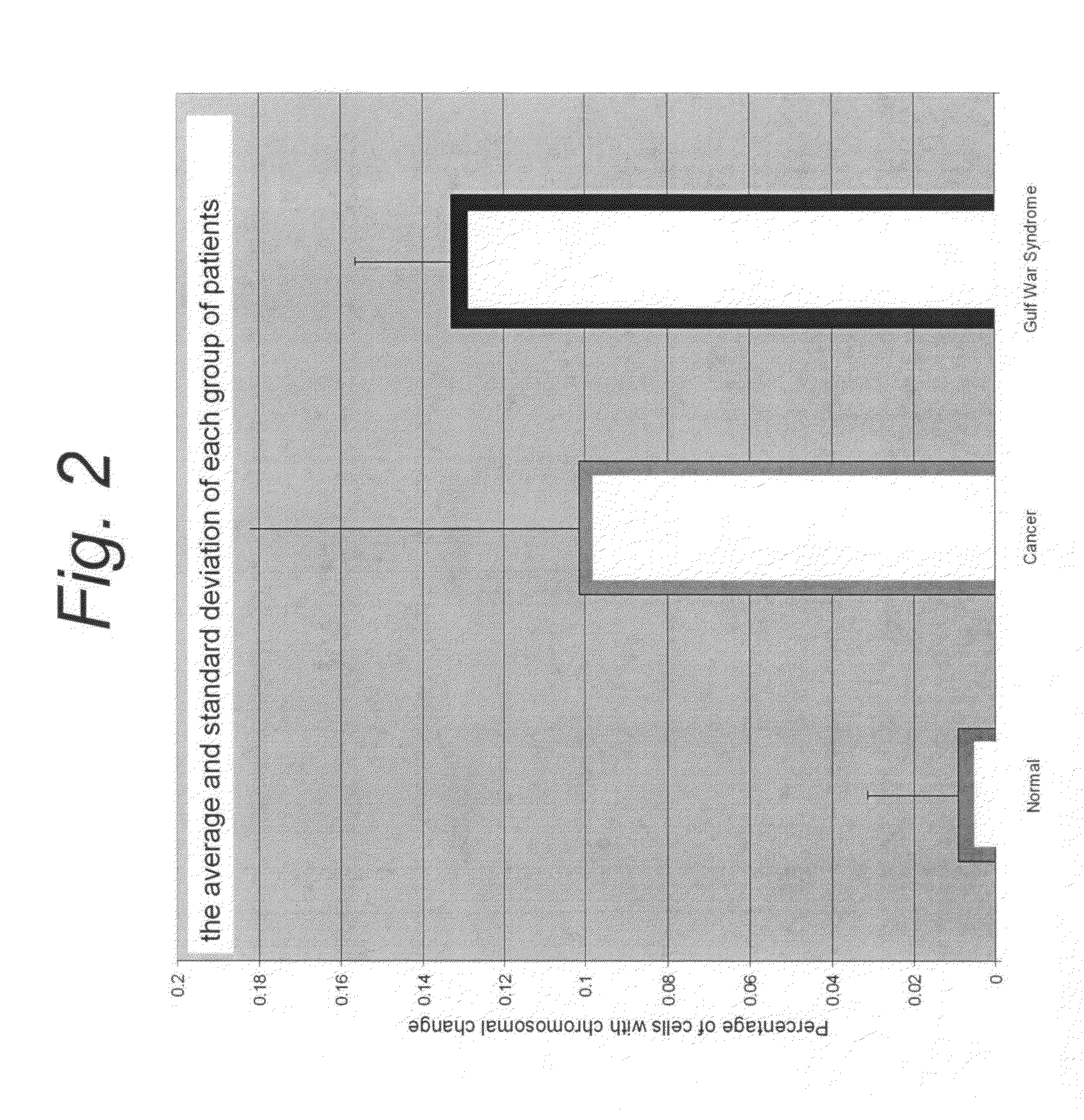
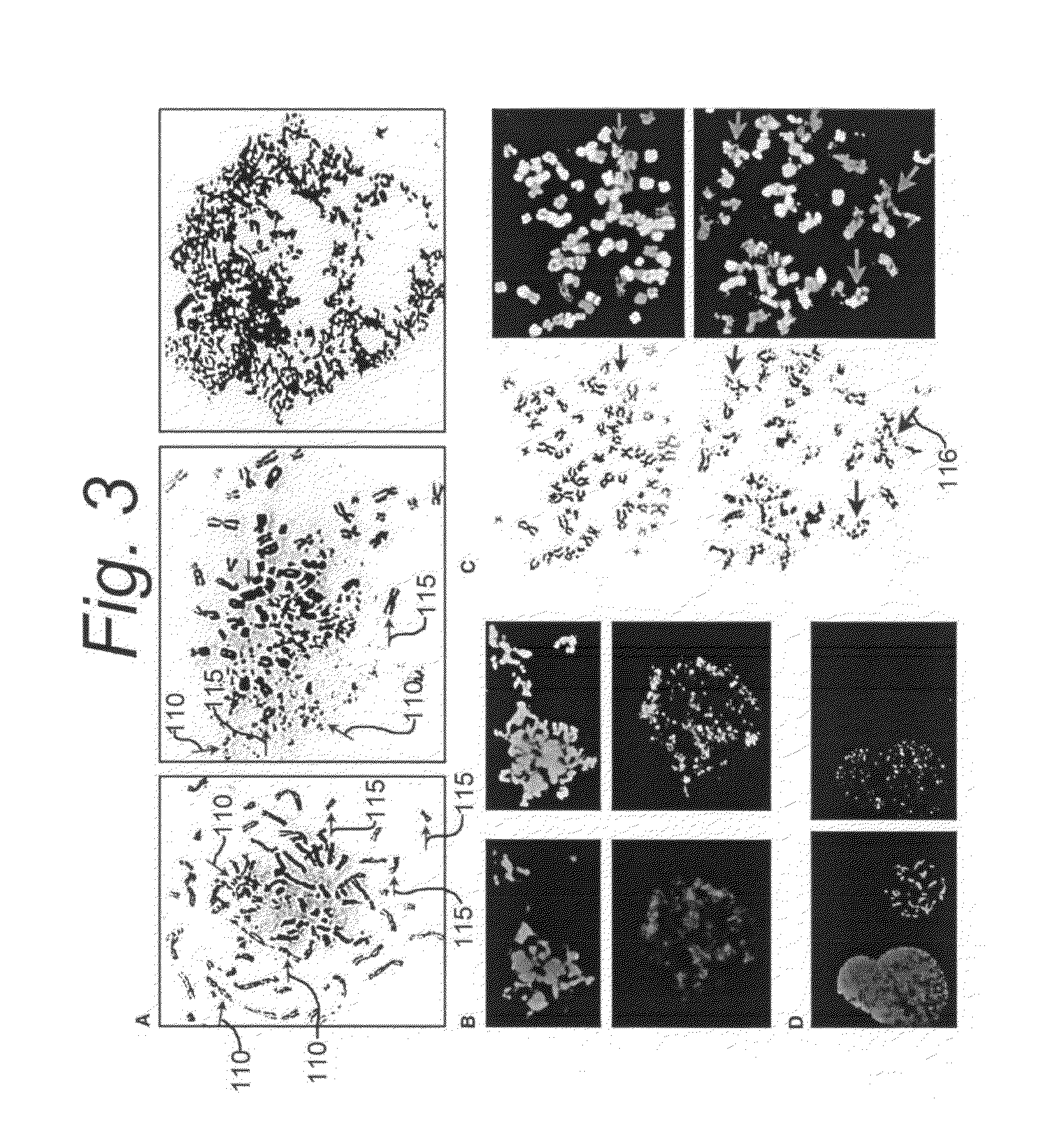
[0105]The following is a detailed description of a new type of NCCA and of a new type of mitotic cell death, termed “chromosome fragmentation,” which is a consequence of certain cellular stressors such as inherited genomic instability or chemotherapeutic treatment in M phase, and a pathologically related process that results in the breakdown of the chromosomes, elimination of genetic material, and subsequent death of cells. This form of cell death is different from typical apoptosis and mitotic catastrophe. It is caspase independent, does not exhibit the typical oligosomal DNA degradation of apoptosis, and is not inhibited by overexpression of Bcl-2.

[0106]Classic methods of inducing mitotic catastrophe do not increase levels of chromosome fragmentation detectable by cytogenetic analysis. Chromosome fragmentation, although morphologically similar to, is distinct from, S-phase premature chromosome condensation (and chromosome pulverization) as chromosomes undergoing fragmentation are ...


PUM

PropertyMeasurementUnit
restrictive temperatureaaaaaaaaaa

Abstract

A diagnostic method of determining tumorigenicity of a tissue specimen includes the steps of determining the magnitude of genome diversity in the tissue specimen, and diagnosing a likelihood of cancer in response thereto. The magnitude of genome diversity includes the determination of karyotypic heterogeneity in tissue specimen, illustratively by detecting non-clonal chromosome aberrations (NCCAs). The detection of NCCAs includes the identification of various types and frequency of NCCAs, and diagnosis is responsive to the step of detecting the frequency of NCCAs. Detection of NCCAs includes the further step of screening lymphocytes. Also, the step of determining the presence of elevated genome diversity includes the step of applying Spectral Karyotyping to detect structural and numerical aberrations throughout the genome. The diagnostic method is useful to determine drug resistance in a patient and potential harmfulness, to evaluate the side effects of drugs, and to measure genome system stress.

Description

RELATIONSHIP TO OTHER APPLICATION(S)[0001]This application is a continuation-in-part patent application of U.S. Ser. No. 12 / 583,194, filed Aug. 14, 2009, which claims the benefit of the filing date of Provisional Patent Application Ser. No. 61 / 188,916, filed on Aug. 14, 2008, the disclosures of which are incorporated herein by reference.GOVERNMENT RIGHTS[0002]This invention was made in part under contract awarded by National Cancer Institute—National Institute of Health, Contract Number R01-CA100247. The government may have certain rights in the invention.BACKGROUND OF THE INVENTION[0003]1. Field of the Invention[0004]This invention relates generally to diagnostic or evaluation methods of detecting the characteristics of cancer or and other complex diseases, and more particularly, to a diagnostic methodology for determining the likelihood of the presence of cancer, or of developing cancer, or of monitoring chemotherapy, or evaluating effectiveness or side effects of drugs, in respon...

Claims


Application Information

Patent Timeline
Patent Type & Authority Applications(United States)
IPC IPC(8): C12Q1/68
CPCC12Q2600/106C12Q1/6886
Inventor HENG, HENRY H.YE A/K/A JING YE, CHRISTINE
Owner HENG HENRY H
Why Eureka
Try Eureka