Image processing device, imaging device, computer-readable recording medium, and image processing method

a technology of imaging device and image processing method, which is applied in the direction of color signal processing circuit, television system, instruments, etc., can solve the problem of difficult to distinguish between normal body tissue and abnormal tissu

Active Publication Date: 2010-09-30
OLYMPUS CORP
View PDF2 Cites 58 Cited by
  • Summary
  • Abstract
  • Description
  • Claims
  • Application Information

AI Technical Summary

Benefits of technology

[0011]An image processing device according to an aspect of the present invention includes a normal-light-image storage unit that stores therein each picture signal of a normal light image of an observed region, the normal light image being captured by a normal-light imaging unit that receives normal light from the observed region via a plurality of color filters having different spectral characteristics. The device also includes a fluorescence-image storage unit that stores therein each picture signal of a fluorescence image of the observed region, the fluorescence image being captured by a fluorescence imaging unit that receives fluorescence generated from the observed region irradiated with excitation light. The device ...

Problems solved by technology

The light-dark contrast of fluorescence makes it difficult to distinguish between a norma...

Method used


Image

  • Image processing device, imaging device, computer-readable recording medium, and image processing method
  • Image processing device, imaging device, computer-readable recording medium, and image processing method
  • Image processing device, imaging device, computer-readable recording medium, and image processing method

Examples


first embodiment

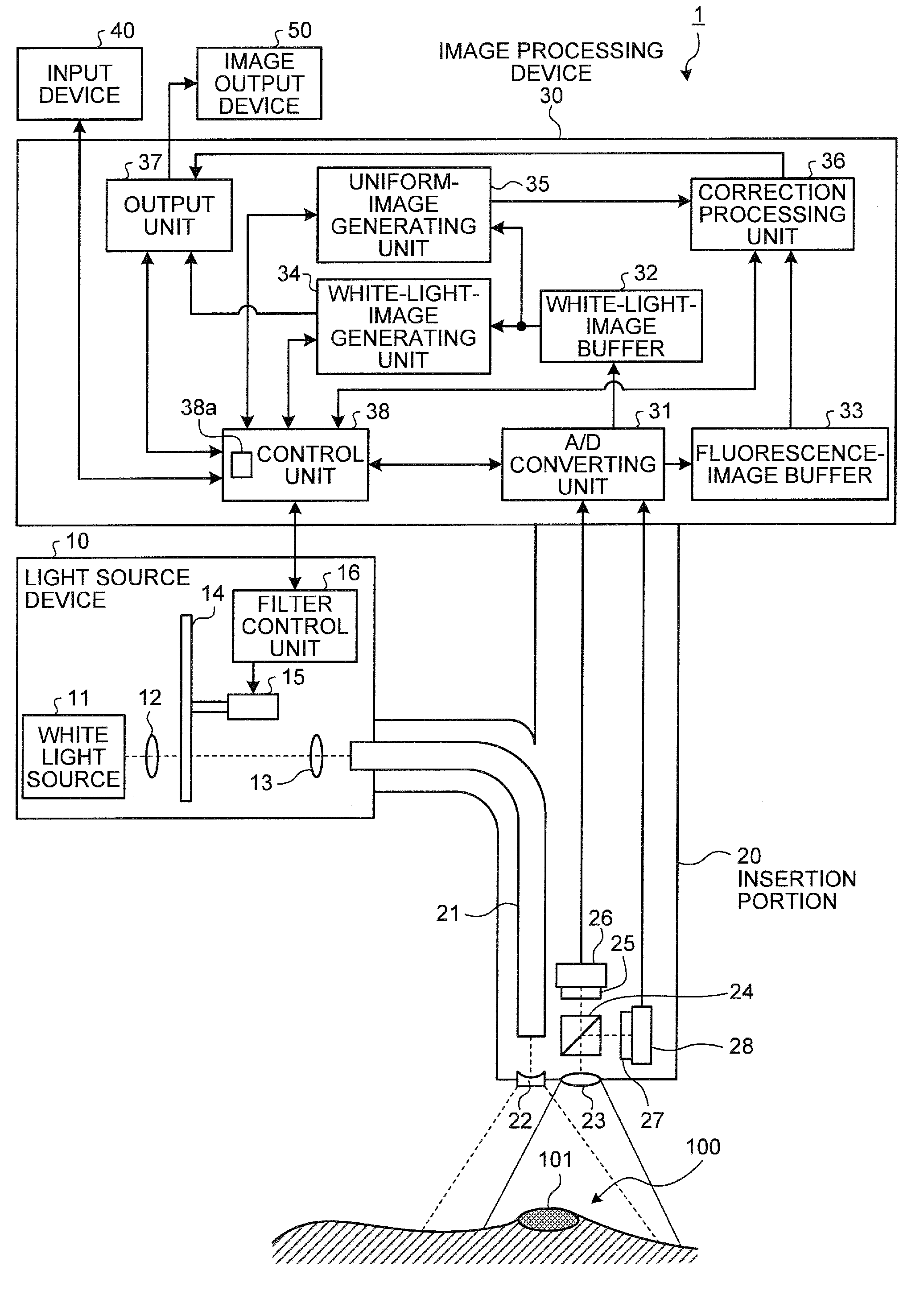
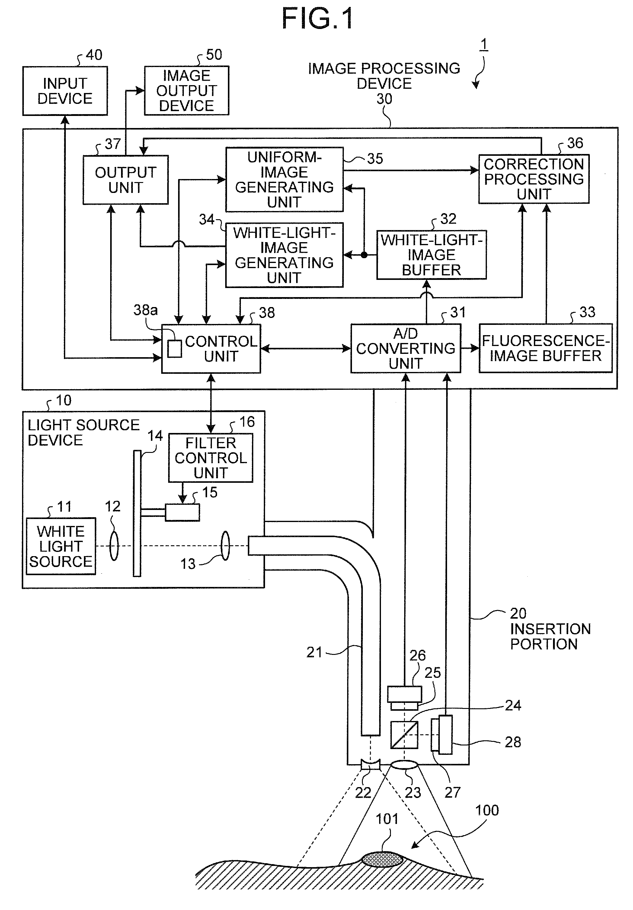
[0041]FIG. 1 is a block diagram schematically showing a configuration example of an endoscope apparatus according to a first embodiment of the present invention. An endoscope apparatus 1 according to the first embodiment is one example of the imaging device of the present invention. As illustrated in FIG. 1, the endoscope apparatus 1 includes a light source device 10 that applies light to an observed region 100 inside a subject, and an elongated insertion portion 20 to be inserted into a body cavity of the subject. The endoscope apparatus 1 also includes an image processing device 30 that processes images of the observed region 100, an input device 40 that inputs various types of information to the image processing device 30, and an image output device 50 that outputs image information processed by the image processing device 30.

[0042]The light source device 10 functions as a light source unit that switchably applies excitation light, which excites a fluorescent agent, and white lig...

second embodiment

[0123]A second embodiment of the present invention will be described. In the first embodiment described above, the uniform image is generated based on the R picture signals corresponding to the red light filter in the color filter group 25, as the image information for correcting the fluorescence image of the observed region 100. However, in the second embodiment, a uniform image is generated based on a picture signal of an infrared component (hereinafter, referred to as “IR picture signal”) corresponding to an infrared light filter in a color filter group arranged on a light receiving portion of a white-light imaging unit.

[0124]FIG. 15 is a block diagram schematically showing a configuration example of an endoscope apparatus according to the second embodiment of the present invention. As illustrated in FIG. 15, an endoscope apparatus 2 according to the second embodiment includes a light source device 210 instead of the light source device 10 of the endoscope apparatus 1 of the firs...

third embodiment

[0160]A third embodiment of the present invention will be described below. In the first and the second embodiments described above, the endoscope apparatuses 1 and 2 are described as examples of the imaging device of the present invention. However, in the third embodiment, a microscope apparatus is described as an example of the imaging device of the present invention, and descriptions of an image processing device, a computer-readable recording medium recording an image processing program, and an image processing method used by the microscope apparatus will be given subsequently.

[0161]FIG. 24 is a block diagram schematically showing a configuration example of the microscope apparatus according to the third embodiment of the present invention. As illustrated in FIG. 24, a microscope apparatus 3 of the third embodiment is an example of the imaging device of the present invention, and includes a microscope body 320 instead of the insertion portion 20 of the endoscope apparatus 1 of th...


PUM


Abstract

An image processing device captures a normal light image of an observed region via a plurality of color filters having different spectral characteristics, respectively, generates a uniform image having low contrast by extracting each picture signal corresponding to a picture signal in a wavelength band for which light absorption characteristic of a contrast region in the observed region becomes low, and corrects each picture signal of a fluorescence image of the observed region by using the uniform image.

Description

CROSS-REFERENCE TO RELATED APPLICATIONS[0001]This application is based upon and claims the benefit of priority from Japanese Patent Application No. 2009-077563, filed on Mar. 26, 2009, the entire contents of which are incorporated herein by reference.BACKGROUND OF THE INVENTION[0002]1. Field of the Invention[0003]The present invention relates to an image processing device, an imaging device, a computer-readable recording medium recording an image processing program, and an image processing method, and particularly relates to an image processing device, an imaging device, a computer-readable recording medium recording an image processing program, and an image processing method for processing a fluorescence image based on fluorescence from a subject.[0004]2. Description of the Related Art[0005]Conventionally, endoscope apparatuses have been used in the medical field to observe inside of organs of a subject. The endoscope apparatuses generally capture a white light image of a body tiss...

Claims


Application Information

Patent Timeline
IPC IPC(8): H04N9/73G06K9/00
CPCA61B1/0638G01N21/645A61B1/0646A61B1/043A61B1/00009A61B1/000094A61B1/0655
Inventor YOSHINO, KOICHIROSUZUKI
Owner OLYMPUS CORP
Why Eureka
Try Eureka