Methods for Image Analysis and Visualization of Medical Image Data Suitable for Use in Assessing Tissue Ablation and Systems and Methods for Controlling Tissue Ablation Using Same

a technology of image data and image analysis, applied in the field of data analysis and visualization techniques, can solve the problem of difficulty in segmentation of medical images

Inactive Publication Date: 2010-10-21
TYCO HEALTHCARE GRP LP
View PDF51 Cites 77 Cited by
  • Summary
  • Abstract
  • Description
  • Claims
  • Application Information

AI Technical Summary

Problems solved by technology

Despite the existence of numerous image segmentation techniques, segmentation of medical images is still a challenge due to the variety and complexity of medical images.

Method used


Image

  • Methods for Image Analysis and Visualization of Medical Image Data Suitable for Use in Assessing Tissue Ablation and Systems and Methods for Controlling Tissue Ablation Using Same
  • Methods for Image Analysis and Visualization of Medical Image Data Suitable for Use in Assessing Tissue Ablation and Systems and Methods for Controlling Tissue Ablation Using Same
  • Methods for Image Analysis and Visualization of Medical Image Data Suitable for Use in Assessing Tissue Ablation and Systems and Methods for Controlling Tissue Ablation Using Same

Examples


Embodiment Construction

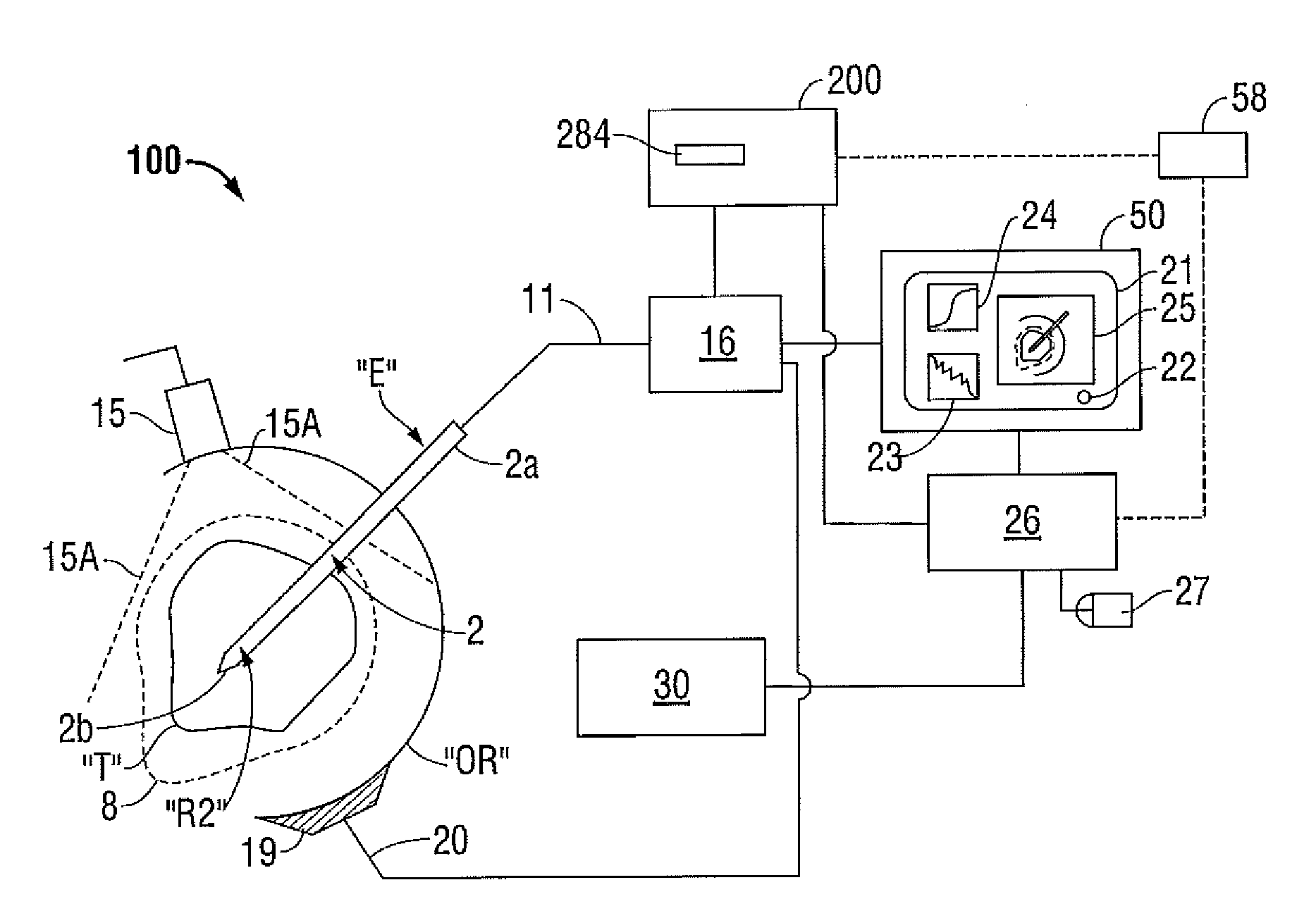
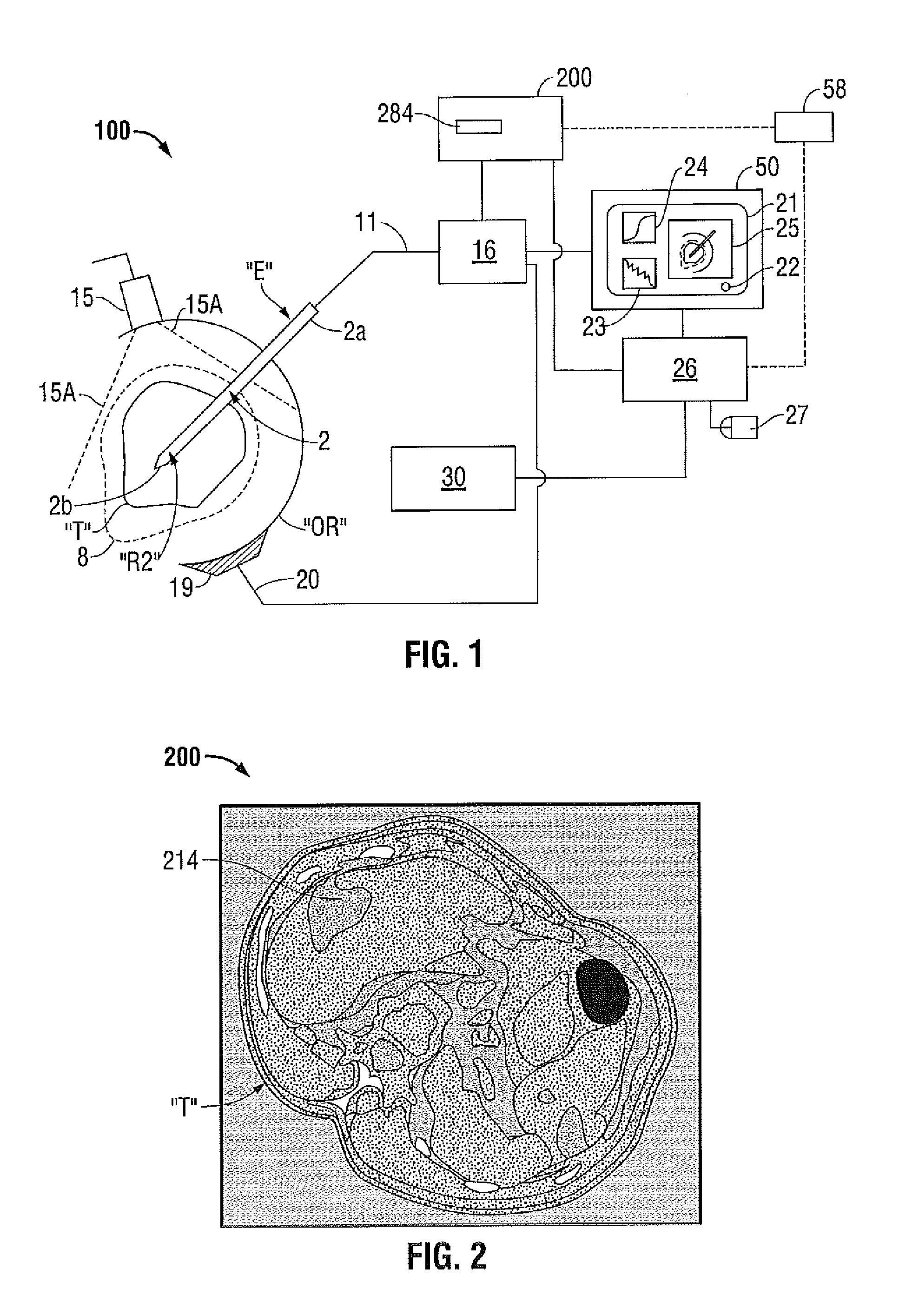
[0036]Hereinafter, embodiments of presently disclosed methods for image analysis and visualization of medical image data and the presently disclosed systems and methods for controlling tissue ablation using the same are described with reference to the accompanying drawings. Like reference numerals may refer to similar or identical elements throughout the description of the figures. As shown in the drawings and as used in this description, and as is traditional when referring to relative positioning on an object, the term “proximal” refers to that portion of the object that is closer to the user and the term “distal” refers to that portion of the object that is farther from the user.

[0037]This description may use the phrases “in an embodiment,”“in embodiments,”“in some embodiments,” or “in other embodiments,” which may each refer to one or more of the same or different embodiments in accordance with the present disclosure. For the purposes of this description, a phrase in the form “A...


PUM


Abstract

A method of image analysis includes the initial step of receiving a data set including image data. The image data represents a sequence of 2-D slice images. The method includes the steps of segmenting an object of interest from surrounding image data of each slice image based on a p-value of a t-statistic relating each pixel successively examined to statistical properties derived from pixel values within the region of interest, and rendering a volume of the object of interest using (x,y) coordinates corresponding to boundaries of the segmented object of interest.

Description

CROSS-REFERENCE TO RELATED APPLICATION[0001]The present application is a continuation-in-part application, which claims priority to, and the benefit of, U.S. patent application Ser. No. 12 / 761,267 filed on Apr. 15, 2010, which claims priority to, and the benefit of, U.S. Provisional Application Ser. No. 61 / 169,556 filed on Apr. 15, 2009, the disclosures of which are herein incorporated by reference in their entireties.BACKGROUND[0002]1. Technical Field[0003]The present disclosure relates to data analysis and visualization techniques, and, more particularly, to methods for image analysis and visualization of medical image data that are suitable for use in assessing biological tissue ablation, and systems and methods for controlling tissue ablation using the same.[0004]2. Discussion of Related Art[0005]Treatment of certain diseases requires the destruction of malignant tissue growths, e.g., tumors. Electromagnetic radiation can be used to heat and destroy tumor cells. Treatment may in...

Claims


Application Information

Patent Timeline
Patent Type & Authority Applications(United States)
IPC IPC(8): A61B18/14G06K9/34
CPCA61B18/1477A61B18/18A61B19/50A61B19/52A61B2018/1869G06T2207/30096G06T7/0081G06T7/0087G06T15/08G06T2207/10072G06T2207/20076A61B2019/5276A61B90/36A61B34/10A61B2090/378G06T7/11G06T7/143
Inventor COE, JONATHAN A.LATDKOW, CASEY M.
Owner TYCO HEALTHCARE GRP LP
Why Eureka
Try Eureka