Method and system for performing upright magnetic resonance imaging of various anatomical and physiological conditions

Inactive Publication Date: 2011-01-13
FONAR
View PDF2 Cites 5 Cited by
  • Summary
  • Abstract
  • Description
  • Claims
  • Application Information

AI Technical Summary

Benefits of technology

[0014]The present invention is not limited only to the use of MR signal intensity to perform the methods described herein. In other another aspects of the present invention, any type of MR signal characteristic (e.g. signal

Problems solved by technology

Also, MRI can obtain images of soft tissues within the body which are not readily visualized using other imaging techniques.
This method o

Method used


Image

  • Method and system for performing upright magnetic resonance imaging of various anatomical and physiological conditions
  • Method and system for performing upright magnetic resonance imaging of various anatomical and physiological conditions
  • Method and system for performing upright magnetic resonance imaging of various anatomical and physiological conditions

Examples


examples

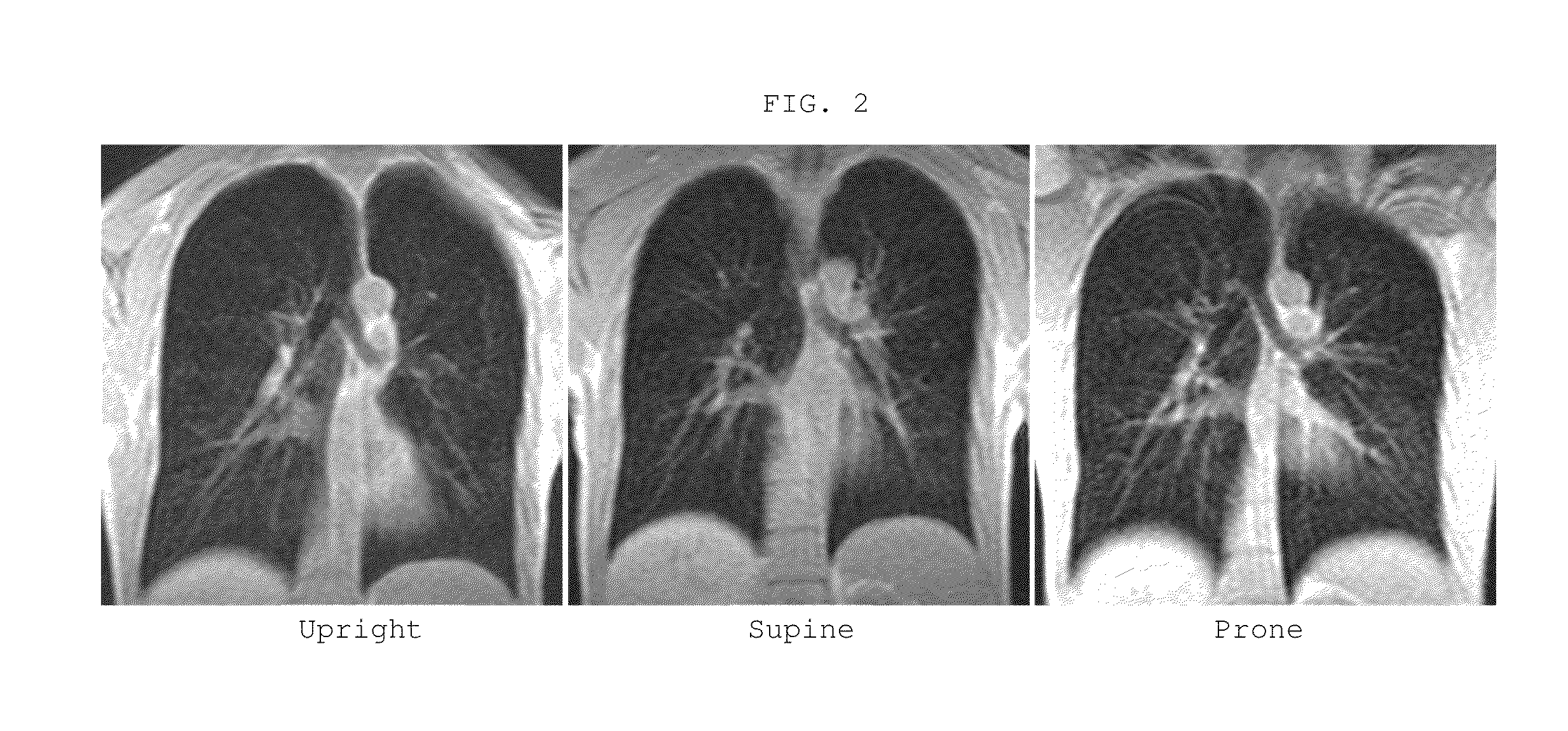
[0060]The following example describes a study performed using upright lung MR imaging on four volunteer subjects.

[0061]Scans were performed on the UPRIGHT® MRI scanner (Fonar Corporation, New York) at 0.6 T. With a vertical walk-in patient space, the scanner has a bed that can be rotated to any angle between the vertical and horizontal position. As a result, the patient can be scanned standing up, sitting up, flexing, extending, and lying horizontally or in the reverse-Trendelenburg position. When combined with various patient orientations such as feet first head last, Trendelenburg and lateral decubitus positions are also possible.

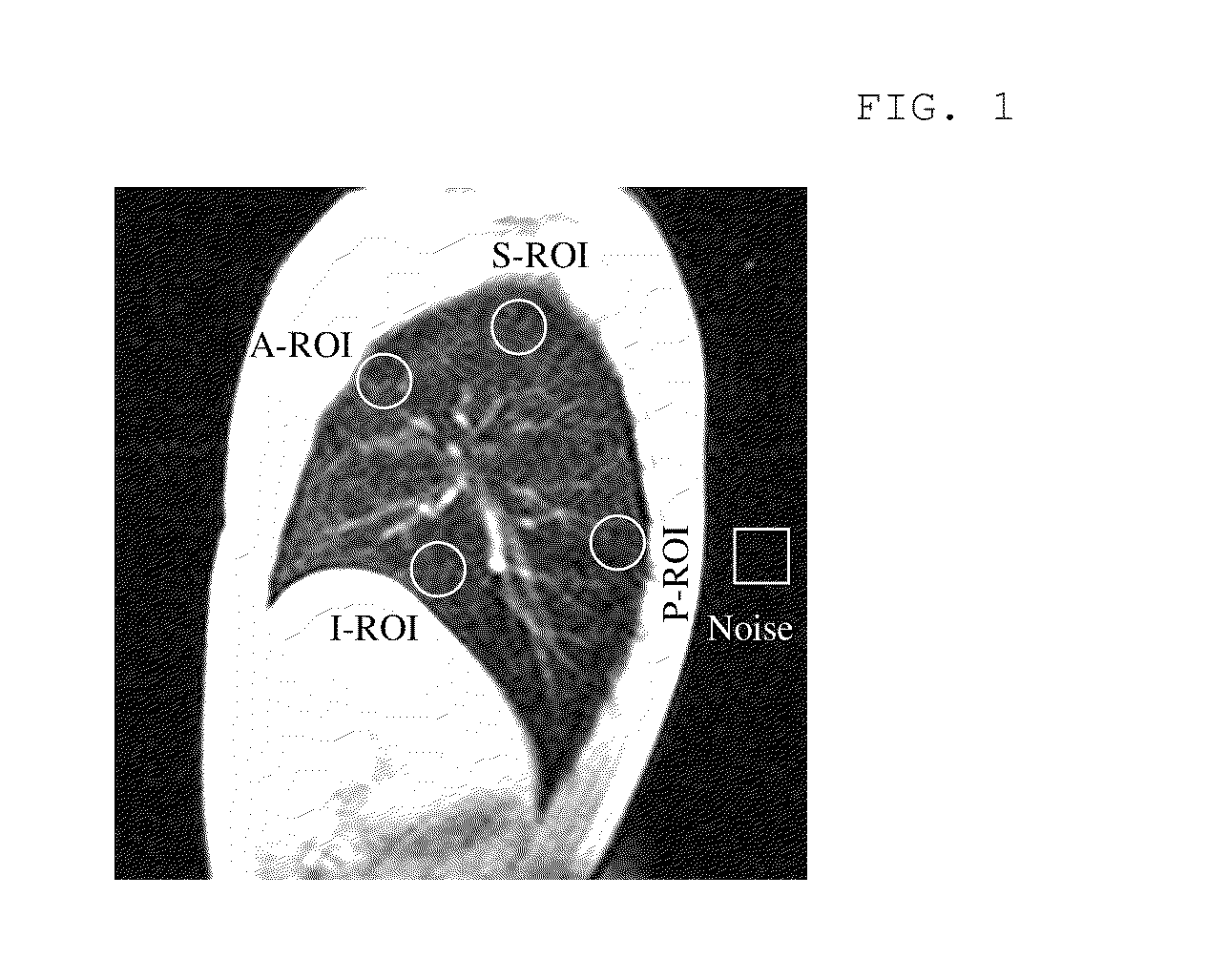
[0062]Four non-smoking healthy human volunteers participated in this study (2 males / 2 females, age: 22-54). A body RF transmitter coil was used for RF excitation. A separate rigid thoracic coil (quadrature receive-only) with a homogenous illumination was centered on the lungs. The imaging parameters were selected according to clinical parameters commonly ...


PUM


Abstract

Vasculature or parenchyma is imaged using upright MRI techniques, on patients who may have conditions such as congestive heart failure, or otherwise be healthy. When an individual is horizontal, venous drainage is minimized, causing the vessels to remain engorged, also referred to herein as vascular congestion. Vascular congestion results in an enlarging of the vessels and surrounding tissue causing the vessels to be more visible on MRIs. The decrease in vascular visibility in upright subjects is in part, due to an increase in venous drainage. Patients suffering from coronary and/or pulmonary deficiencies (e.g. CHF) experience decreased rates and degrees of venous drainage. In one embodiment, the present invention uses upright imaging to visualize these enlarged vessels.

Description

CROSS-REFERENCE TO RELATED APPLICATIONS[0001]This application claims the benefit of the filing date of U.S. Provisional Patent Application No. 61 / 270,405 filed Jul. 8, 2009, the disclosure of which is hereby incorporated herein by reference.BACKGROUND[0002]Magnetic resonance imaging (“MRI”) offers numerous advantages over other imaging techniques. MRI does not expose either the patient or medical personnel to X-rays and offers important safety advantages. Also, MRI can obtain images of soft tissues within the body which are not readily visualized using other imaging techniques. This feature makes MRI particularly useful in analyzing the parenchymal lung tissue and pulmonary vasculature in certain patients.[0003]Different tissues produce different signal characteristics. Tissues having a high density of nuclei will produce stronger signals than tissues with a low density of such nuclei. Furthermore, relatively small gradients in the magnetic field are superimposed on the static magne...

Claims


Application Information

Patent Timeline
IPC IPC(8): A61B5/055
CPCA61B5/055A61B5/416A61B5/08A61B5/0044A61B5/0263
Inventor DAMADIAN, RAYMOND V.CHU, KI-CHEUNG
Owner FONAR
Why Eureka
Try Eureka