System for Medical Image Processing, Manipulation and Display

a medical image and user interface technology, applied in the field of xray medical image user interface system, can solve the problems of cumbersome manipulation, user limited options to view a part, and additional time-consuming steps

Active Publication Date: 2011-03-10
SIEMENS HEALTHCARE GMBH
View PDF18 Cites 27 Cited by
  • Summary
  • Abstract
  • Description
  • Claims
  • Application Information

AI Technical Summary

Benefits of technology

[0004]A system provides a secondary display of an enlarged region of interest, automatically selected in response to collimator position and a detected (or user selected) object position, on a secondary live display while concurrently presenting an overview X-ray image on a primary live display. An X-ray medical image user interface system includes one or more displays for displaying medical images. At least one display concurrently presents, a first image window including an X-ray image overview of a portion of patient anatomy and a second image window

Problems solved by technology

A user has limited options to view a part of an image in detail and enlarged while still using X-ray imaging in providing an overv

Method used


Image

  • System for Medical Image Processing, Manipulation and Display
  • System for Medical Image Processing, Manipulation and Display
  • System for Medical Image Processing, Manipulation and Display

Examples


Embodiment Construction

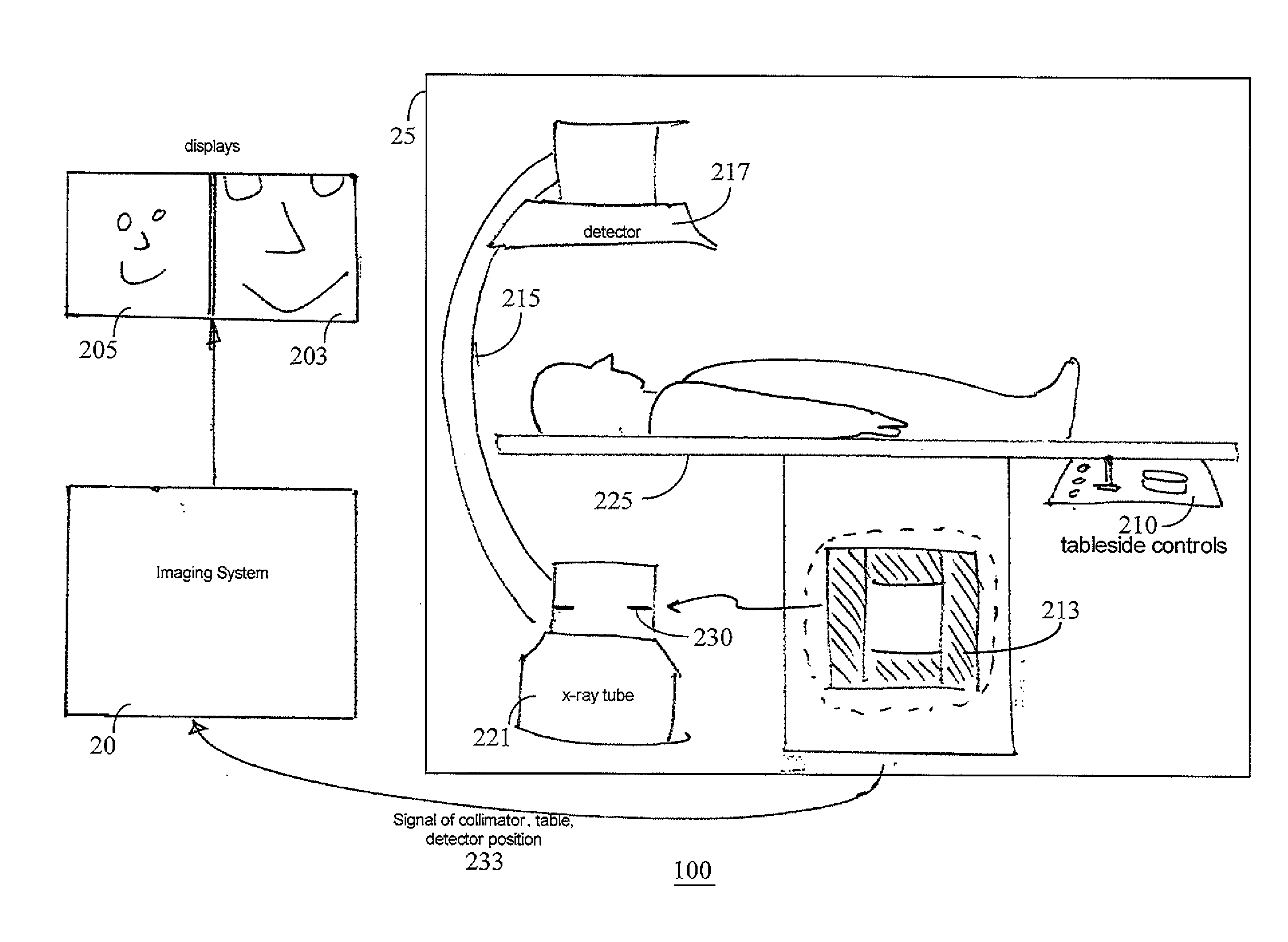
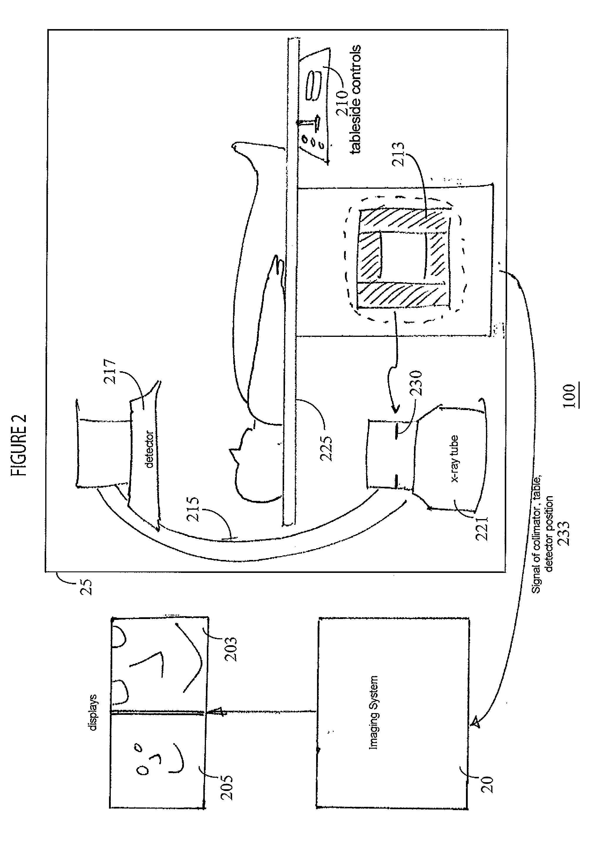
[0014]A system according to invention principles provides a secondary display of an enlarged region of interest on a secondary live display while concurrently presenting an overview X-ray image on a primary live display. The enlarged region of interest is automatically selected in response to collimator position.

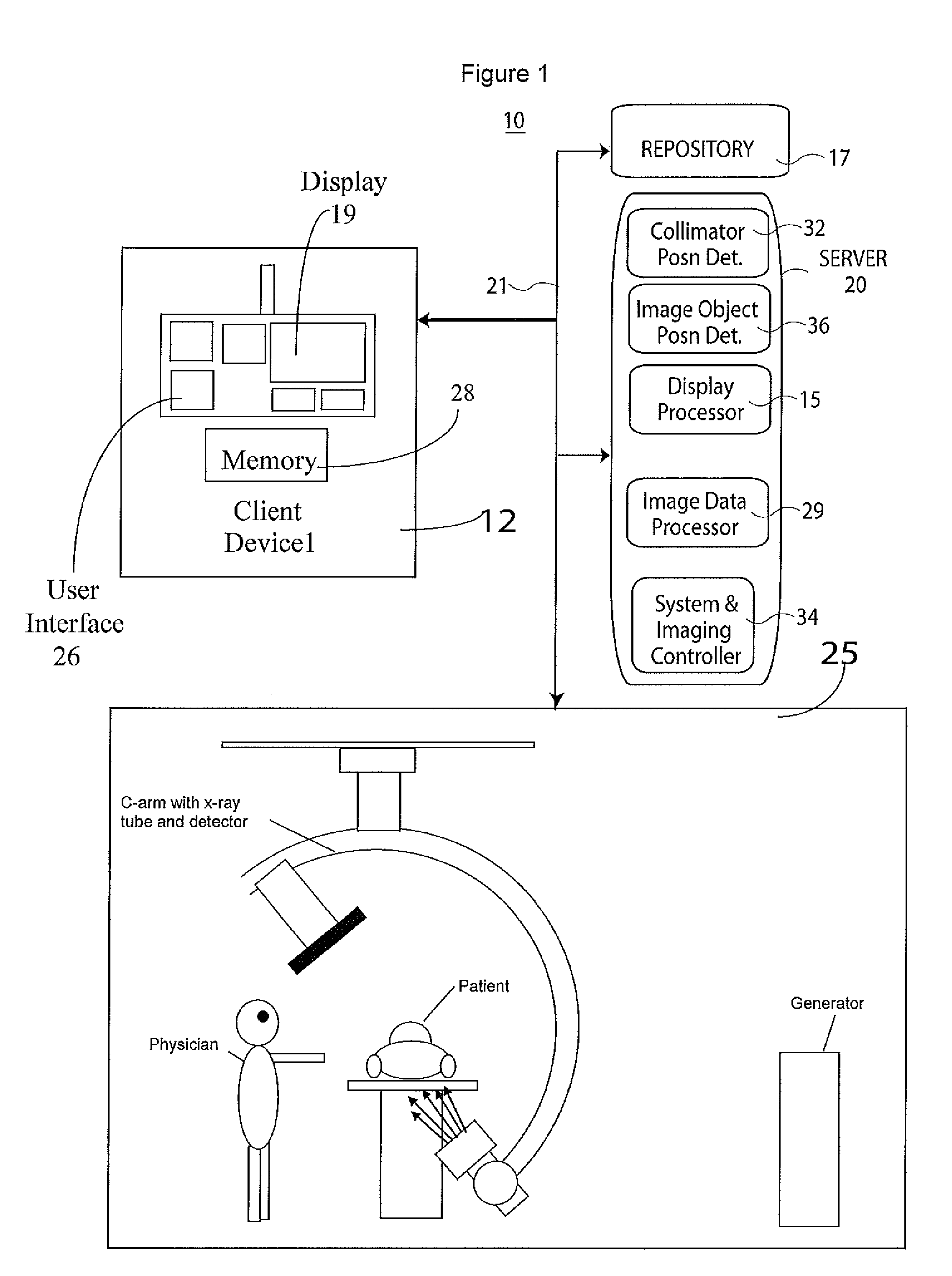
[0015]FIG. 1 shows an X-ray medical image user interface system 10 including one or more displays for displaying medical images. System 10 includes one or more processing devices (e.g., computers, workstations or portable devices such as notebooks, Personal Digital Assistants, phones) 12 that individually include a user interface control device 26 such as a keyboard, mouse, touchscreen, voice data entry and interpretation device, at least one display 19 and memory 28. System 10 also includes at least one repository 17, X-ray imaging modality system 25 (which in an alternative embodiment may comprise an MR (magnetic resonance), CT scan, or Ultra-sound system, for example) and...


PUM


Abstract

A system automatically adaptively adjusts an enlarged region of interest presented as a zoomed image on a secondary live display in response to X-ray filter (e.g., collimator) adjustment. An X-ray medical image user interface system includes one or more displays for displaying medical images. At least one display concurrently presents, a first image window including an X-ray image overview of a portion of patient anatomy and a second image window including an enlarged region of interest within the X-ray image overview. A collimator position detector provides a collimator signal in response to a detected collimator position. An image data processor automatically identifies the enlarged region of interest within the X-ray image overview in response to the collimator signal. A display processor generates data representing an image comprising the enlarged region of interest of the X-ray image overview in response to the identification of the enlarged region of interest within the X-ray image overview.

Description

[0001]This is a non-provisional application of provisional application Ser. No. 61 / 239,824 filed 4 Sep., 2009, by J. Baumgart et al.FIELD OF THE INVENTION[0002]This invention concerns an X-ray medical image user interface system including one or more displays for displaying medical images and an image depicting an enlarged region of interest within an X-ray image.BACKGROUND OF THE INVENTION[0003]In known X-ray systems, during an x-ray procedure, typically only one field of view of a portion of patient anatomy is supported at a time. A user has limited options to view a part of an image in detail and enlarged while still using X-ray imaging in providing an overview of the patient. These options include, after image acquisition, selecting a post processing zoom and pan function to manipulate the image to see a region of interest (ROI), then return to the X-ray image of the overview of the region of interest. These options may also include, changing the field of view by changing the zo...

Claims


Application Information

Patent Timeline
IPC IPC(8): H05G1/64
CPCG06T19/00G06T2219/028
Inventor BAUMGART, JOHNDEVADAS, BENITA
Owner SIEMENS HEALTHCARE GMBH
Why Eureka
Try Eureka