Computer readable medium, systems and methods for improving medical image quality using motion information

a motion information and computer-readable medium technology, applied in image enhancement, instruments, computing, etc., can solve the problems of scan device noise, detract from image quality, and motion analysis techniques that have not been widely exploited in clinical settings

Inactive Publication Date: 2011-03-31
QI IMAGING
View PDF14 Cites 3 Cited by
  • Summary
  • Abstract
  • Description
  • Claims
  • Application Information

AI Technical Summary

Problems solved by technology

Noise from the scanning device may also detract from image quality.
While motion analysis techniques have been used to interpolate between regularly sampled video frames, motion analysis techniques have not been widely exploited in the clinical setting.

Method used


Image

  • Computer readable medium, systems and methods for improving medical image quality using motion information
  • Computer readable medium, systems and methods for improving medical image quality using motion information
  • Computer readable medium, systems and methods for improving medical image quality using motion information

Examples


Embodiment Construction

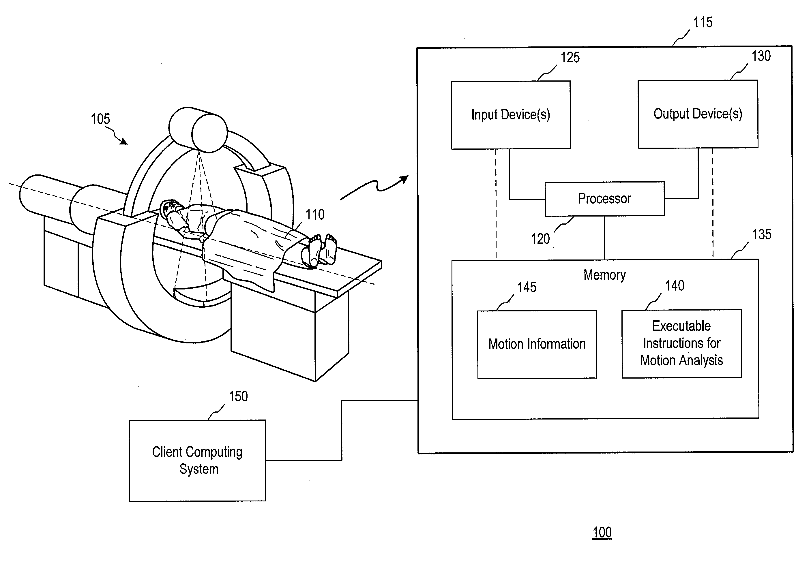
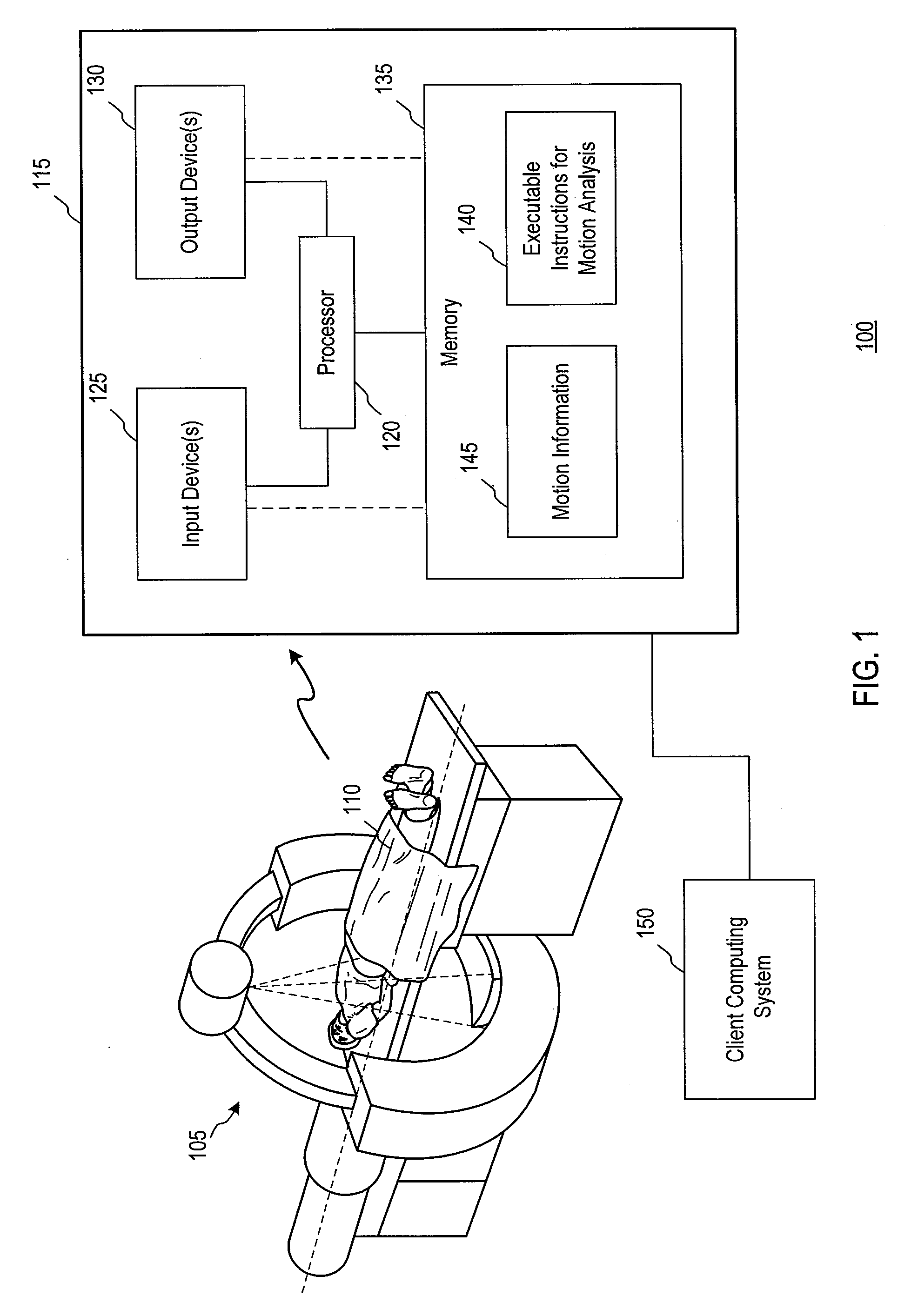
[0017]FIG. 1 is a schematic illustration of a medical scenario 100 in accordance with an embodiment of the invention. A computed tomography (CT) scanner 105 is shown and may collect data from a subject 110. The data may be transmitted to an imaging system 115 for processing. The imaging system 115 may include a processor 120, input devices 125, output devices 130, a memory 135, or combinations thereof As will be described further below, the memory 135 may store executable instructions for performing motion analysis 140. Following the processing of volume data using motion analysis, motion information 145 may be stored in the memory 135. The motion information 145 may be used in a variety of ways, as will be described further below, to generate or alter volume data that may be displayed on one or more of the output devices 130 or transmitted for display by a client computing system 150. The client computing system 150 may communicate with the imaging system 115 through any mechanism,...


PUM


Abstract

Motion information generated by comparing one or more instances of clinical volume data may be used in a variety of applications. Examples of applications described herein include filtering volume data and adjusting voxel intensity based on the motion information. Motion information may also be used to compress volume data. Combinations of these effects may also be achieved.

Description

TECHNICAL FIELD[0001]The invention relates generally to medical image visualization techniques, and more particularly to the use of motion analysis in the visualization of images.BACKGROUND OF THE INVENTION[0002]A variety of medical devices may be used to generate clinical images, including computed tomography (CT) and magnetic resonance imaging (MRI) scanners. These scanners may generate volume data of human anatomy. In this manner, multiple instances of volume data of an anatomical feature may be generated, and may capture movement or other changes of the feature over time.[0003]For example, 3D clinical images may include cardiac scans with scan intervals under a second. In other examples, the 3D scans may include continuous ultrasound scans generating multiple images per second. In this manner, images may be obtained of deformable organs or other features which may change shape from scan-to-scan depending on a variety of variables, such as a patient's posture or breathing pattern...

Claims


Application Information

Patent Timeline
IPC IPC(8): G06K9/00G09G5/02
CPCG06T5/50
Inventor MATSUMOTO, KAZUHIKO
Owner QI IMAGING
Why Eureka
Try Eureka