Lesion area extraction apparatus, method, and program

a technology for extracting apparatus and lesion areas, applied in the field of lesion area extraction apparatus and method, can solve problems such as the degradation of the extraction performance of the lesion area, and achieve the effects of improving the extraction accuracy of the area, reducing the work load of the user, and ensuring the extraction accuracy

Active Publication Date: 2011-03-31
FUJIFILM CORP
View PDF11 Cites 16 Cited by
  • Summary
  • Abstract
  • Description
  • Claims
  • Application Information

AI Technical Summary

Benefits of technology

[0036]According to the lesion area extraction apparatus, method, and program of the present invention, when extracting a lesion area from a medical image, lesion area extraction processing data used for extracting the lesion area from a plurality of lesion area extraction processing data generated in advance based on a lesion description character provided in a radiology report with respect to the lesion area, and the lesion area is extracted suing the determined lesion area extraction processing data. This allows each lesion area to be extracted by making full use of characteristics of each lesion area, whereby area extraction accuracy may be improved.
[0037]Further, the lesion area is extracted using the position information of the lesion area related to a character string of a radiology report, so that the user does not need to directly specify an arbitrary point in the lesion area, whereby the work load of the user may be reduced.
[0038]Further, in the lesion area extraction apparatus, method, and program described above, if a radiology report is displayed, then a character string is specified in the displayed radiology report, lesion area extraction processing data is determined based on the lesion description character of the specified character string, and the lesion area is extracted using the determined lesion area extraction processing data and the position information of the lesion area related to the specified character string, the lesion area corresponding to the lesion description character of the character string may be extracted accurately by making full use of the characteristics of the lesion area. Further, by displaying the extracted lesion area, radiology report generation or observation efficiency may be improved.

Problems solved by technology

Lesion areas, however, differ in characteristics from type to type and the method that extracts all types of lesion areas by the same evaluation function described above does not fully make use of the characteristics of the respective lesion areas, leading to the degradation in extraction performance of a lesion area.

Method used


Image

  • Lesion area extraction apparatus, method, and program
  • Lesion area extraction apparatus, method, and program
  • Lesion area extraction apparatus, method, and program

Examples


Embodiment Construction

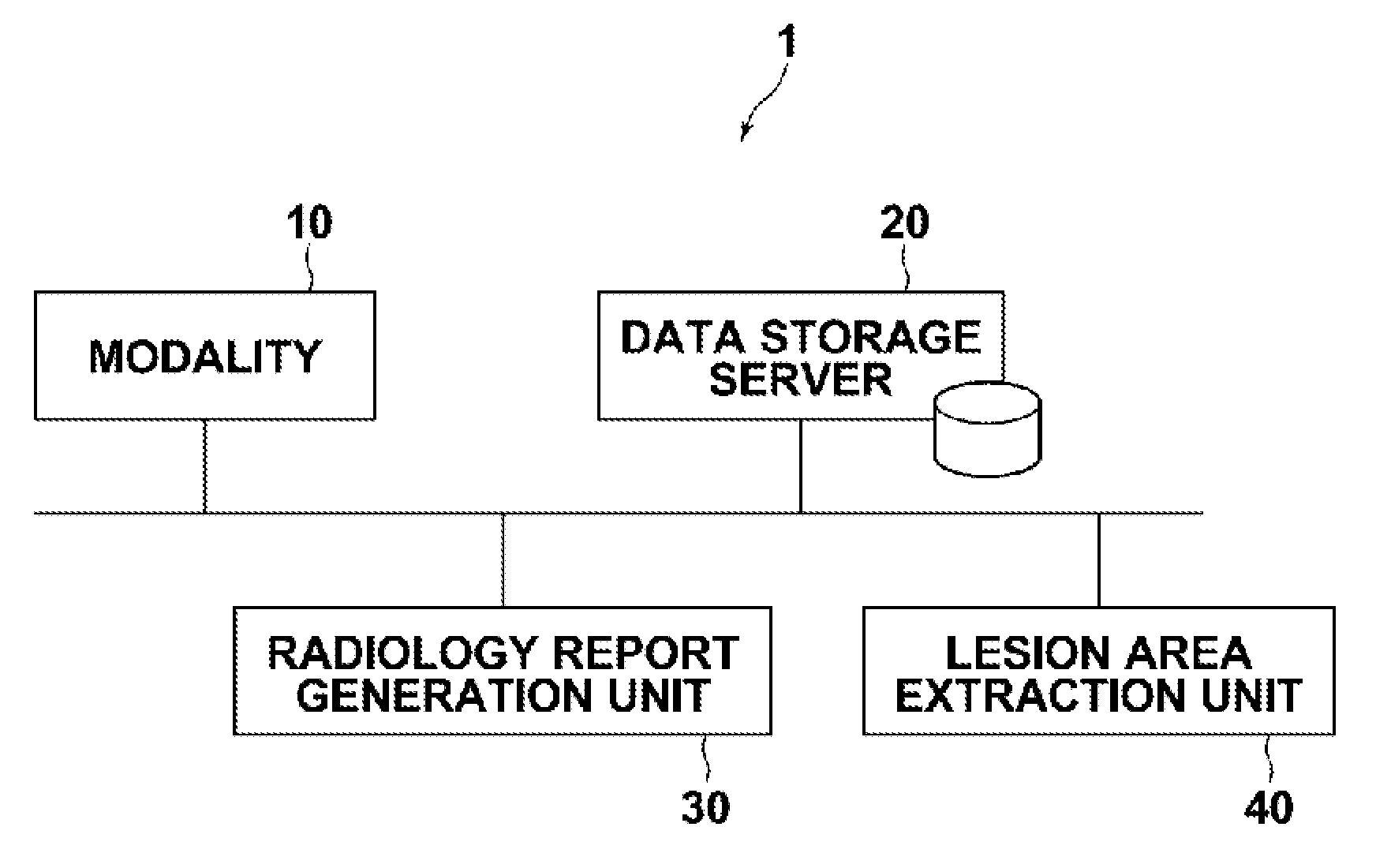
[0046]Hereinafter, embodiments of the present invention will be described with reference to the accompanying drawings. FIG. 1 is a block diagram of a medical image processing system, schematically illustrating the configuration thereof. As shown in FIG. 1, the system includes modality 10, data storage server 20, radiology report generation unit 30, and lesion area extraction unit 40 which are communicatably linked to each other via network 9.

[0047]Modality 10 is a system for generating a medical image representing a test body, and more specifically it is a CT system, MRI system, PET, ultrasonic diagnostic system, or the like. The image generated by modality 10 is sent to data storage server 20 and stored therein.

[0048]Data storage server 20 is a computer for storing / managing various types of data and communicates with other components via network 9 to send / receive image data and the like. More specifically, a medical image generated by modality 10, a radiology report generated by ra...


PUM


Abstract

Recording a plurality of lesion area extraction processing data generated in advance according to a plurality of types of lesion areas, recording a radiology which includes a character string having a lesion description character and being related to position information of a lesion area in the medical image, determining lesion area extraction processing data used for the extraction from the plurality of lesion area extraction processing data based on the lesion description character provided in the radiology report, and performing the extraction using the determined lesion area extraction processing data and the position information of the lesion area related to the character string.

Description

BACKGROUND OF THE INVENTION[0001]1. Field of the Invention[0002]The present invention relates to a lesion area extraction apparatus and method using a radiology report of a medical image.[0003]The invention also relates to a computer readable recording medium on which is recorded a program for causing a computer to perform the method.[0004]2. Description of the Related Art[0005]In medical image diagnosis, it is often the case that image reading is performed by a radiologist and the result of the reading is recorded and reported to the attending doctor as a radiology report, in which electronic radiology reports are widely used.[0006]As one of such radiology reports, a report in which a lesion description character is linked to a reference image that includes the lesion area is known. Use of such radiology report allows the attending doctor to click on the lesion description character in the report displayed on a screen to display and observe a corresponding reference image, thereby ...

Claims


Application Information

Patent Timeline
Patent Type & Authority Applications(United States)
IPC IPC(8): G06K9/00G16H30/40
CPCG06F19/3487G06F19/321G16H15/00G16H30/40
Inventor MORIYA, YOSHIYUKI
Owner FUJIFILM CORP
Why Eureka
Try Eureka