Video adapter for laryngoscope

a technology of video adapter and laryngoscope, which is applied in the field of imaging devices, can solve the problems of not even a good place to position the video monitor, extra minutes and even seconds of delay can be fatal, and the typical medical endoscope is an expensive and bulky instrumen

Inactive Publication Date: 2011-10-06
LASER VADIM
View PDF21 Cites 80 Cited by
  • Summary
  • Abstract
  • Description
  • Claims
  • Application Information

AI Technical Summary

Benefits of technology

[0014]The present invention also provides a swivel unit as a part of the monitor mounting feature for quick directional alignment of the monitor screen.
[0015]The present invention also provides a disposable, inexpensive probe securement or clip that allows the same imaging probe to be used with a variety of existing permanent or disposable laryngoscope blades.

Problems solved by technology

This procedure is not a trivial one especially when it needs to be performed in the inconvenience of emergency type situations and with the patients having various irregularities and complications in the throat and neck areas.
Extra minutes and even seconds of delay can be fatal.
However, a typical medical endoscope is an expensive and bulky instrument, utilizing powerful light sources and fiber optic bundles for delivery of light and / or images.
In such situations, there is not even a good place to position the video monitor for observation of images sent by the endoscope or a camera.
The endoscopes currently known are rather bulky and expensive.
High price and inconvenient construction often preclude use of the endoscopes in the battlefield, on the streets and other ambulance situations.

Method used


Image

  • Video adapter for laryngoscope
  • Video adapter for laryngoscope
  • Video adapter for laryngoscope

Examples


Embodiment Construction

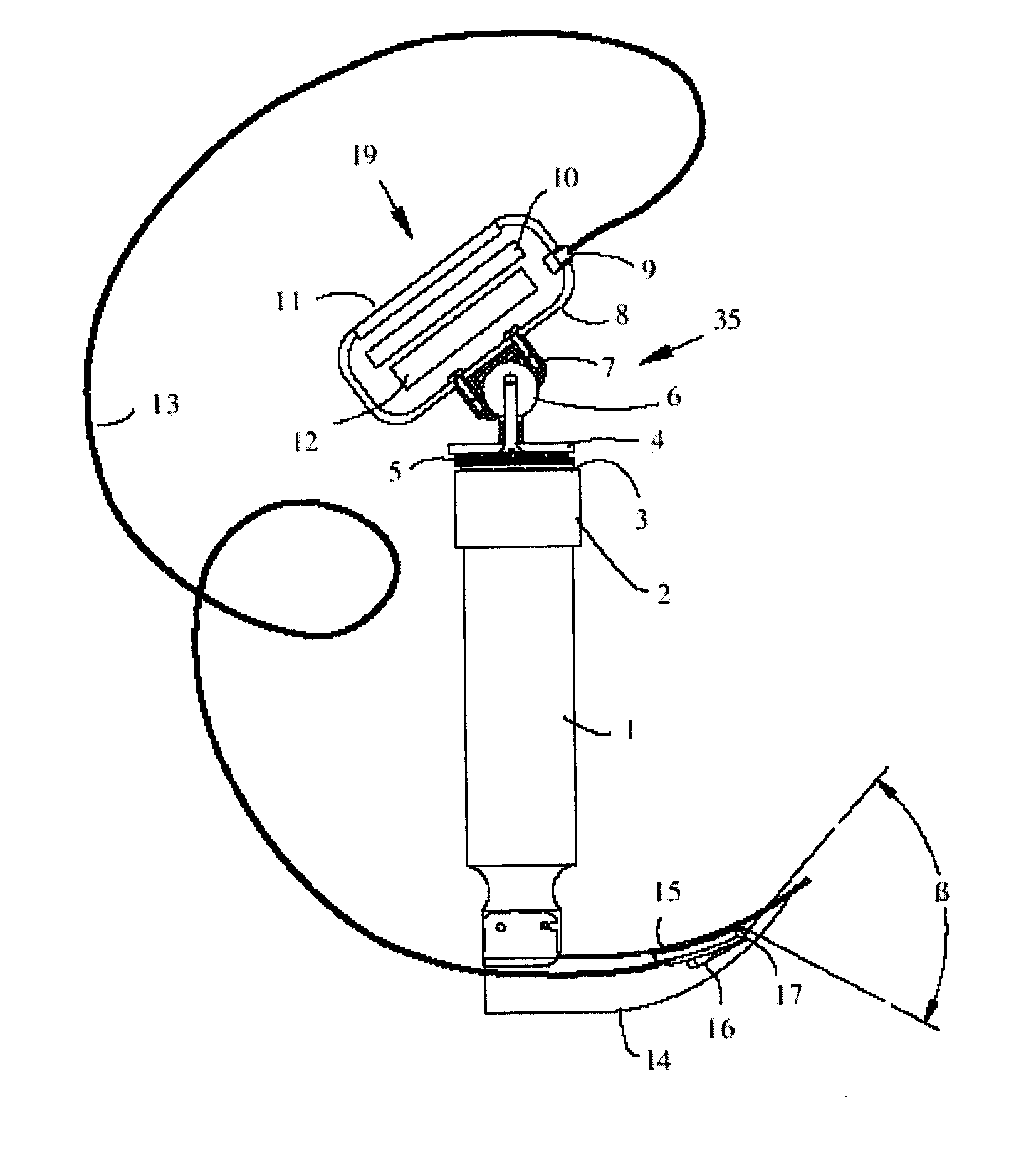
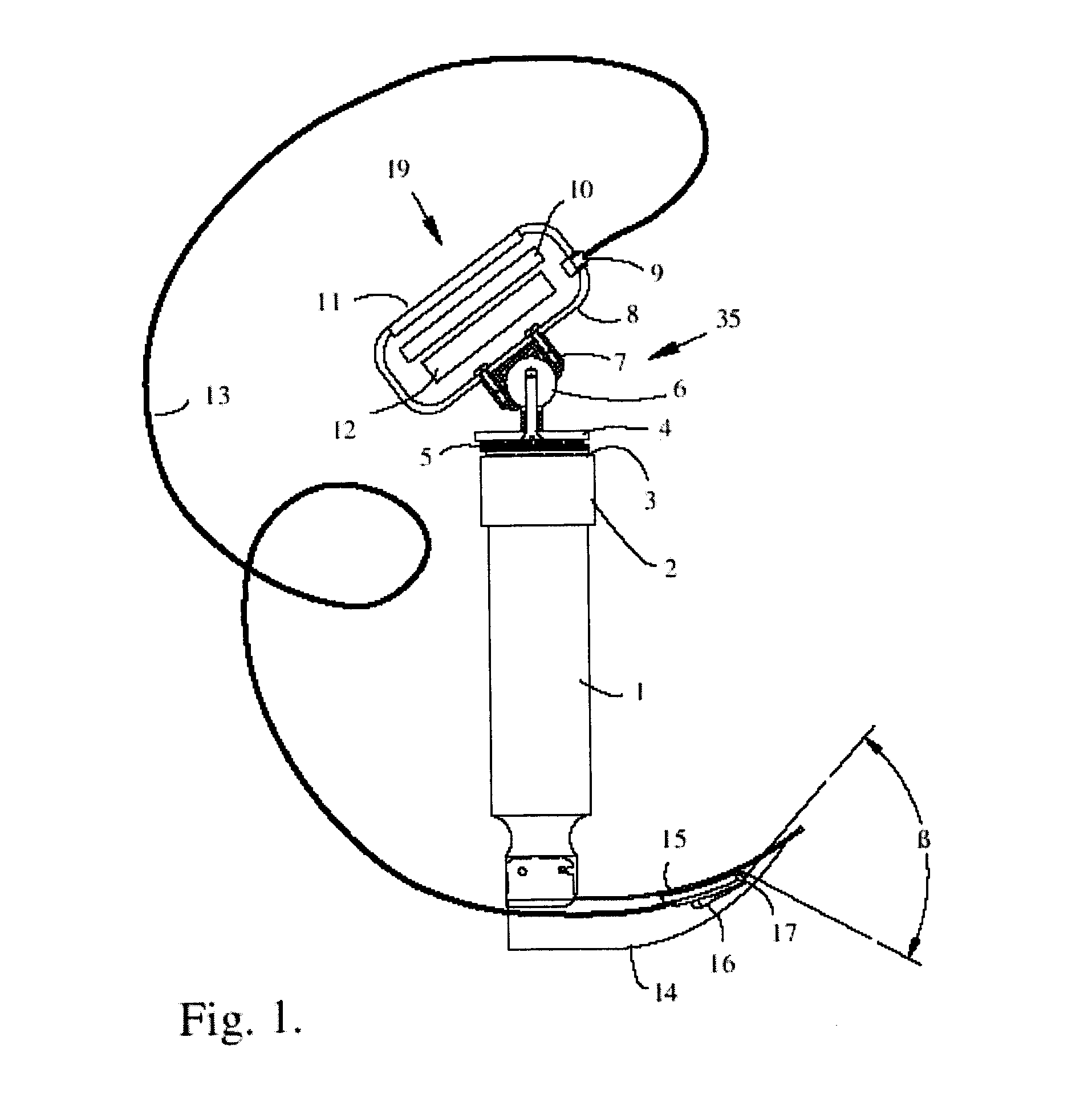
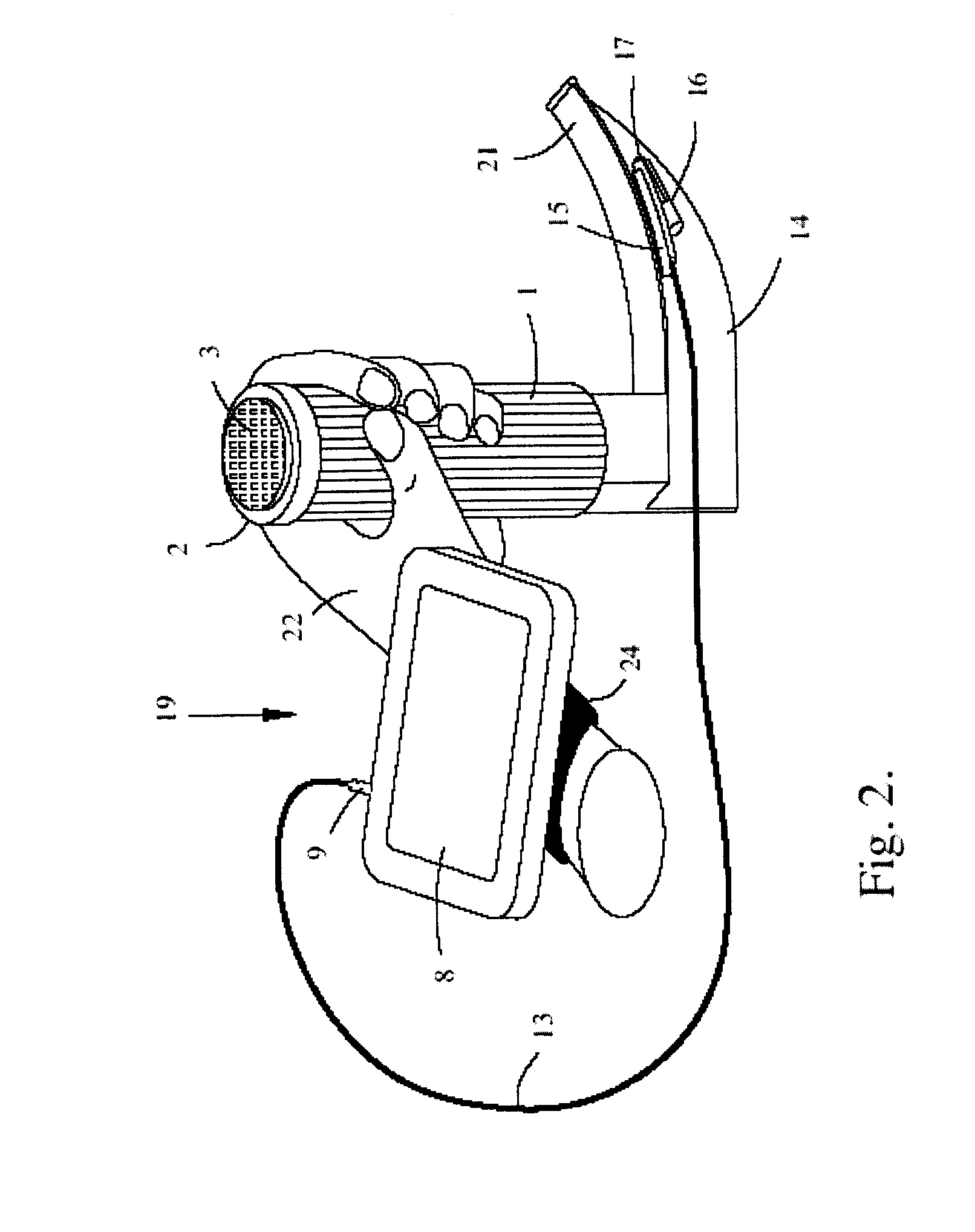
[0029]FIG. 1 illustrates how components of the portable video adapter system are interconnected. The slim endoscope probe 17 is attached to the laryngoscope blade 14 by means of a securement, illustrated as a clip 15, next to an integral light source 16 of the laryngoscope blade. The bracket can include an elongated U-shaped channel having a base and opposed side walls. When the probe is inserted in the channel, it is securely held there by the walls, which normally are inclined towards each other and pushed apart by the body of the probe. Other type of the bracket can include a removable adhesive strip inside the channel. A probe-holding clip can be made as a part of the laryngoscope blade. Depending on type of the laryngoscope, the light source 16 can be either a bulb, or an end of the fiber bundle delivering light from a bulb residing inside the laryngoscope handle 1. A portable video display monitor 19 is provided, mounted on the top of the cup 2 of the laryngoscope handle 1. A ...


PUM


Abstract

A portable, low power, low cost, video adapter for a common laryngoscope, having a slim self-illuminating CMOS camera, attachable with a disposable bracket to the laryngoscope blade and connected via flexible wire to the miniature battery powered color LCD monitor that is detachably mounted either on the wrist band or on top of laryngoscope handle.

Description

CROSS-REFERENCE TO RELATED APPLICATIONS[0001]This application claims the benefit of priority to U.S. provisional application 61 / 318,819, filed Mar. 30, 2010, the disclosure of which is incorporated by reference.FIELD OF THE INVENTION[0002]This invention is concerned with an imaging device used for assisting emergency medical service (EMS) personal and other medical practitioners during performance of an endotracheal intubation procedure.BACKGROUND OF THE INVENTION[0003]Endotracheal intubation is a commonly used procedure during which a semi rigid plastic tube, called an endotracheal tube, is inserted into a patient's trachea through the open mouth. Typically, a laryngoscope is used to view the throat area through the open mouth while the tip of the endotracheal tube advances toward the vocal cords. This procedure is not a trivial one especially when it needs to be performed in the inconvenience of emergency type situations and with the patients having various irregularities and comp...

Claims


Application Information

Patent Timeline
Patent Type & Authority Applications(United States)
IPC IPC(8): A61B1/04
CPCA61B1/00052A61B1/00105A61B1/267A61B1/0676A61B1/0684A61B1/05
Inventor LASER, VADIM
Owner LASER VADIM
Why Eureka
Try Eureka