
Radiological image capturing and displaying method and apparatus

a technology of radiological images and displaying methods, applied in the field of radiological image capturing and displaying methods and apparatuses, can solve the problems of increasing the fatigue of observers, and achieve the effect of reducing the amount of acquired shifts

Inactive Publication Date: 2012-02-02
FUJIFILM CORP
View PDF0 Cites 4 Cited by
  • Summary
  • Abstract
  • Description
  • Claims
  • Application Information

AI Technical Summary

Benefits of technology

The present invention provides a method and apparatus for capturing and displaying a stereo image that can reduce the shift in the image caused by the position of the detector. This is achieved by acquiring the shift amount between the two radiological images and displaying the stereo image using the two images that are relatively shifted in a direction perpendicular to the parallax direction. This can make the stereo image easier for the observer to recognize and reduces fatigue. The invention can be used in a radiological image capturing and displaying apparatus.

Problems solved by technology

As such, when there is a shift between two subject images P1 and P2 forming a stereo image in the Y direction and the observer observes the stereo image displayed on a monitor 3, the burden on the observer to recognize the stereo image increases, which results in an increase in the fatigue of the observer.

Method used


Image

  • Radiological image capturing and displaying method and apparatus
  • Radiological image capturing and displaying method and apparatus
  • Radiological image capturing and displaying method and apparatus

Examples


Embodiment Construction

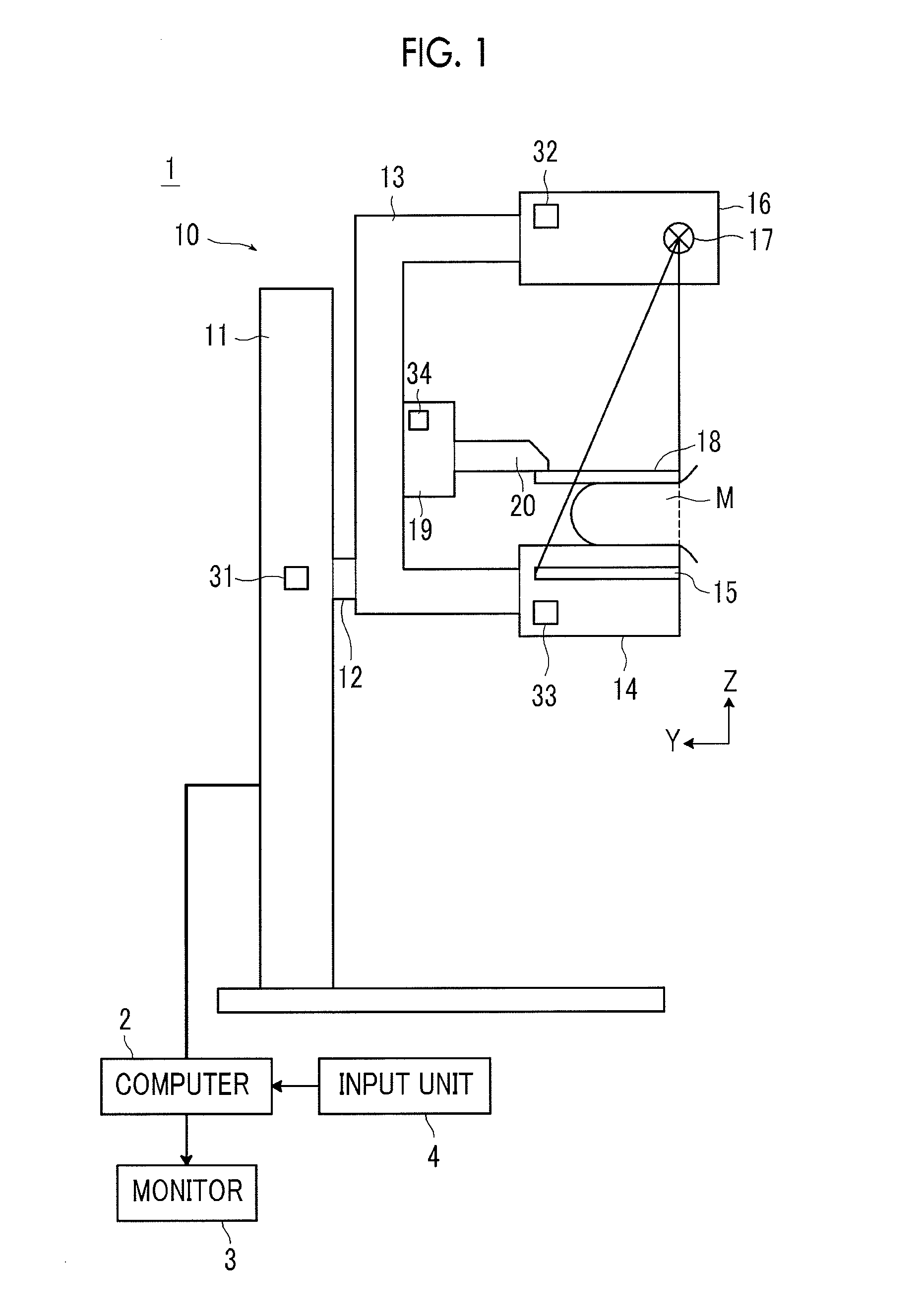
[0039]Hereinafter, a breast image capturing and displaying system using a radiological image capturing and displaying apparatus according to an embodiment of the present invention will be described with reference to the accompanying drawings. FIG. 1 is a diagram schematically illustrating the overall structure of the breast image capturing and displaying system according to this embodiment.

[0040]As shown in FIG. 1, a breast image capturing and displaying system 1 according to this embodiment includes a breast imaging apparatus 10, a computer 2 that is connected to the breast imaging apparatus 10, and a monitor 3 and an input unit 4 that are connected to the computer 2.

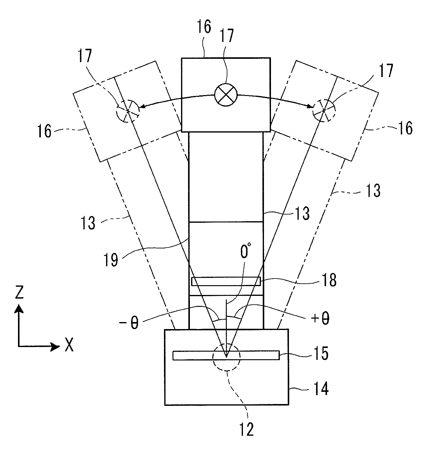
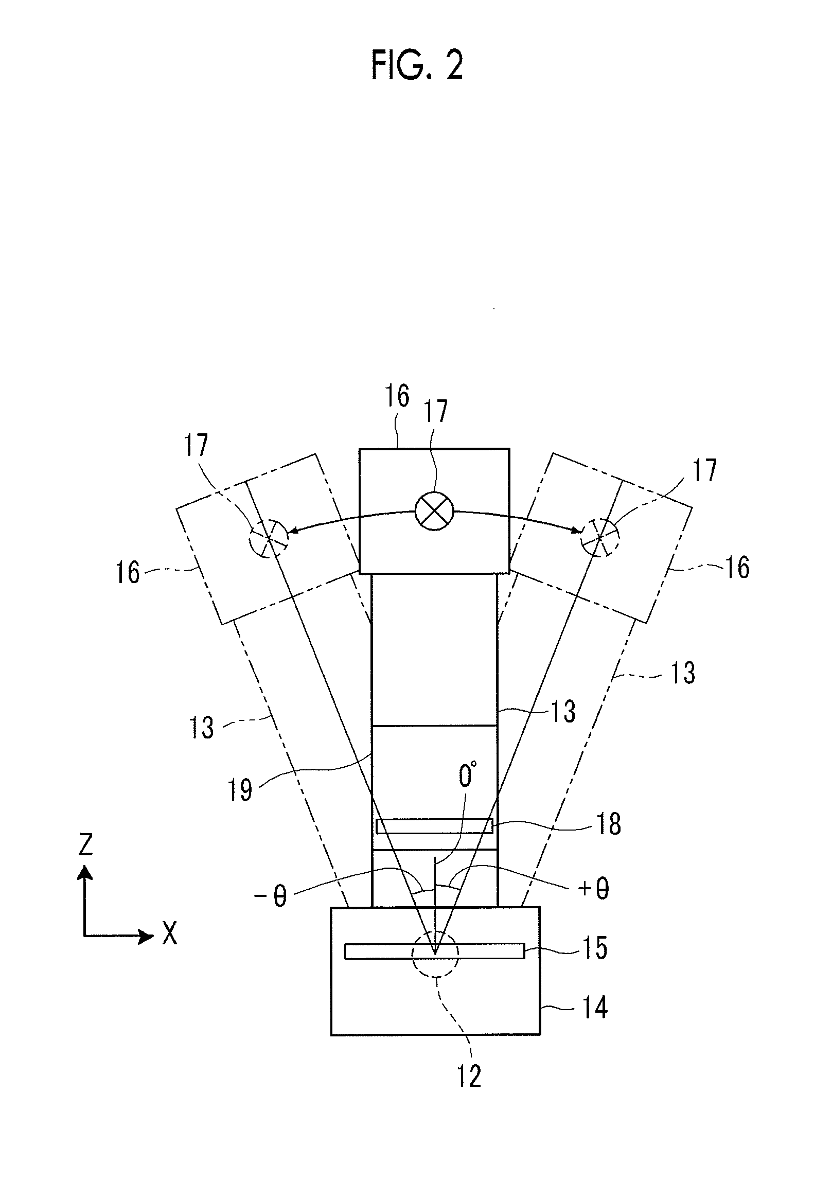
[0041]As shown in FIG. 1, the breast imaging apparatus 10 includes a base 11, a rotating shaft 12 that is movable in the vertical direction (Z direction) relative to the base 11 and is rotatable, and an arm unit 13 that is connected to the base 11 by the rotating shaft 12. FIG. 2 shows the arm unit 13, as viewed from t...


PUM


Abstract

Even when one side of a radiological image detector is inclined with respect to a straight line linking two focal positions of radiation, an appropriate stereoscopic image is displayed. The amount d of shift between two radiological images in a direction perpendicular to a parallax direction is acquired and a stereoscopic image is displayed using the two radiological images which are relatively shifted in the direction perpendicular to the parallax direction so that the acquired amount d of shift is reduced.

Description

BACKGROUND OF THE INVENTION[0001]1. Field of the Invention[0002]The present invention relates to a radiological image capturing and displaying method and apparatus that captures a subject in different radiographing directions to acquire radiological images from the different radiographing directions and displays a stereoscopic image using the radiological images.[0003]2. Description of the Related Art[0004]A technique is known which combines a plurality of images and displays the combined image, thereby enabling stereoscopically viewing using parallax. The image that can be stereoscopically viewed (hereinafter, referred to as a stereoscopic image or a stereo image) is generated based on a plurality of images which are acquired by capturing the same subject from different directions and have parallax therebetween.[0005]Moreover, such way of generating stereoscopic image is utilized not only in the field of digital cameras and televisions but also in the field of capturing a stereosco...

Claims


Application Information

Patent Timeline
Patent Type & Authority Applications(United States)
IPC IPC(8): A61B6/02
CPCA61B6/022A61B6/0414A61B6/5235A61B6/502A61B6/4435
Inventor NAKAYAMA, HIROKI
Owner FUJIFILM CORP
Learn More