Method and apparatus for automatic verification of endotracheal intubation

a technology of endotracheal intubation and automatic verification, which is applied in the field of endotracheal intubation, can solve the problems of unfavorable esophageal intubation, unfavorable esophageal intubation, and inability to obtain a clear view of the entrance to the trachea,

Inactive Publication Date: 2012-05-10
TUBEEYE MEDICAL
View PDF4 Cites 16 Cited by
  • Summary
  • Abstract
  • Description
  • Claims
  • Application Information

AI Technical Summary

Benefits of technology

[0023]Further, in accordance with an embodiment of the invention, the progression of anatomical features is at least one of a cari...

Problems solved by technology

Unfortunately, it's not always possible to obtain a clear view of the entrance to the trachea.
In such cases there is a substantial risk of inserting the ETT into the esophagus instead of the trachea.
Unintentional esophageal intubation can produce catastrophic results as the patient might be deprived of oxygen.
If an ETT remains for too long in the esophagus, it may result in brain damage and even death.
Furthermore, even though the tube might be confirmed to be in an air conducting structure, it may not be correctly positioned.
Prolonged single lung ventilation might...

Method used


Image

  • Method and apparatus for automatic verification of endotracheal intubation
  • Method and apparatus for automatic verification of endotracheal intubation
  • Method and apparatus for automatic verification of endotracheal intubation

Examples


Embodiment Construction

[0040]In the following detailed description, numerous specific details are set forth in order to provide a thorough understanding of the invention. However, it will be understood by those skilled in the art that the invention may be practiced without these specific details. In other instances, well-known methods, procedures, and components have not been described in detail so as not to obscure the invention.

[0041]As described in the Background, the prior art comprises a variety of tools and methods for the insertion of an ETT, verifying its correct placement and monitoring its operation over time. However, whether used individually or in combination, these tools and methods may suffer from a variety of drawbacks. For example, they are often expensive, cumbersome and / or may rely on the presence of an expert operator. Verification of correct placement may be unreliable or untimely, with possibly catastrophic results for the patient.

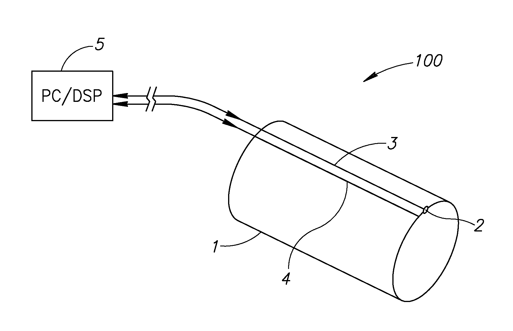
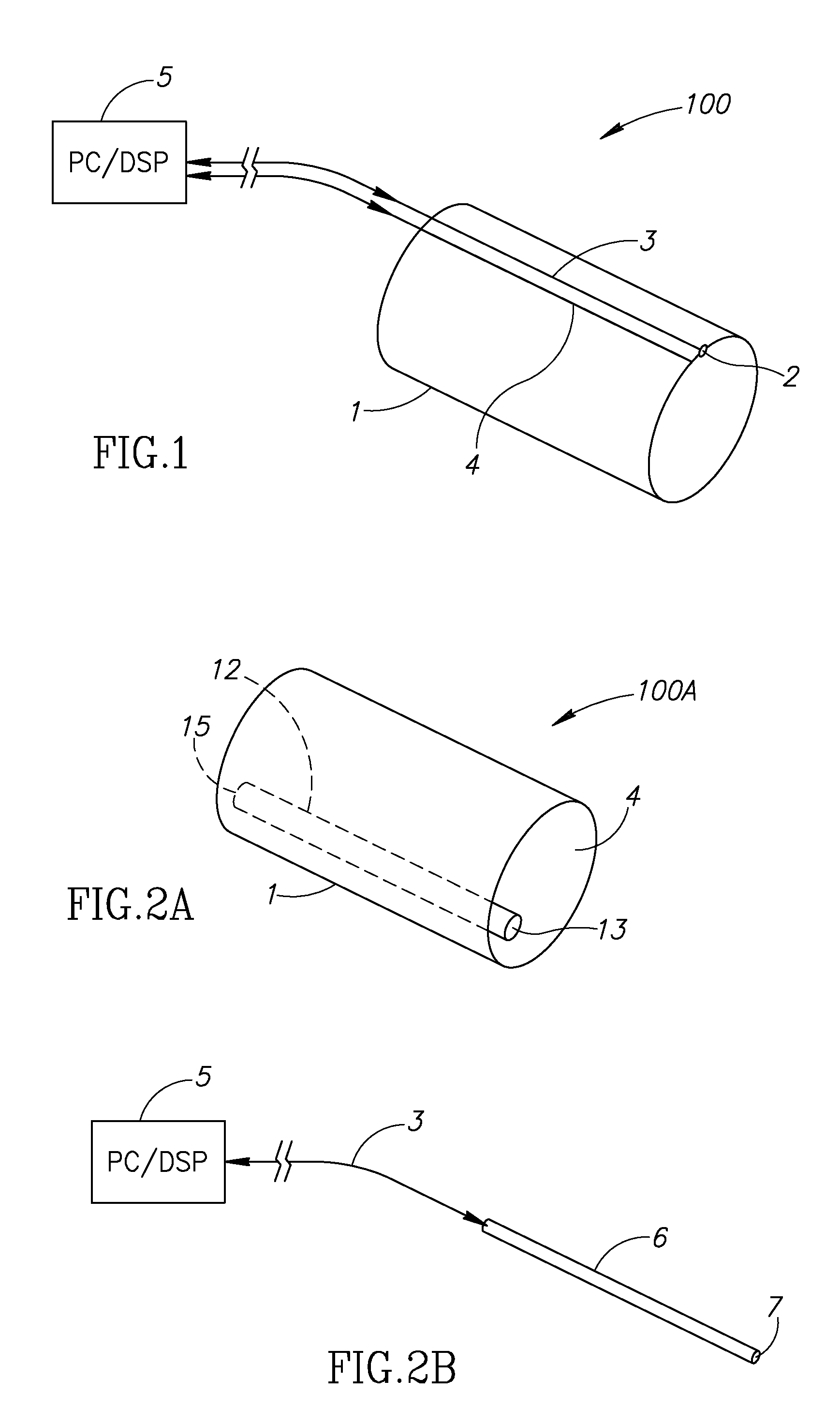
[0042]Applicants have realized that by adding visuali...


PUM


Abstract

A medical device includes a tube, at least one imaging sensor coupled to an endoscope in the tube, and a monitor application to monitor positioning of the tube in a medical patient by identifying expected anatomical features in images provided by the at least one sensor. A method for endotracheal intubation includes receiving imaging frames from a sensor located in an endotracheal tube inserted through a patient's mouth, and processing the imaging frames to identify a progression of anatomical features consistent with a proper placement of the endotracheal tube.

Description

CROSS-REFERENCE TO RELATED APPLICATIONS[0001]This application claims benefit from U.S. Provisional Patent Application No. 61 / 173,324, filed Apr. 28, 2009, which is hereby incorporated in its entirety by reference.FIELD OF THE INVENTION[0002]The present invention relates to medical intubation devices generally and to endotracheal intubation in particular.BACKGROUND OF THE INVENTION[0003]Endotracheal tubes (ETTs) are known in the art. An ETT is a ventilating tube, typically made of plastic, which is introduced into a patient's trachea by a medical procedure known as “intubation”. ETTs ensure the patency (unblocked state) of the patient's airways by creating a safe artificial corridor for air to enter the lungs without obstruction.[0004]To place the ETT in the trachea, an operator typically uses a tool, such as a laryngoscope, to move the tongue out of the way and illuminate the back of the throat. The laryngoscope is inserted into the patient's mouth and passes over the tongue, thusly...

Claims


Application Information

Patent Timeline
IPC IPC(8): A61B1/267A61B1/04A61B1/07A61M1/00A61B1/307A61B1/273A61B1/233A61M16/04A61B1/00
CPCA61B1/05A61B1/267A61M16/0411A61M16/0488A61M2205/50A61M16/04A61M16/0427A61B1/000094A61B1/00055
Inventor LEDERMAN, DROR
Owner TUBEEYE MEDICAL
Why Eureka
Try Eureka