Method and system for segmenting a liver object in an image

a liver object and image technology, applied in image analysis, image enhancement, instruments, etc., can solve the problems of large inter- and intra-expert variations, inability to produce sufficient accurate results, time-consuming manual segmentation of liver structures in ct images, etc., to facilitate subsequent processing steps

Inactive Publication Date: 2012-08-16
AGENCY FOR SCI TECH & RES
View PDF10 Cites 27 Cited by
  • Summary
  • Abstract
  • Description
  • Claims
  • Application Information

AI Technical Summary

Benefits of technology

[0006]In general terms, the present invention proposes in a first aspect a segmentation technique having a quality which is improved by identifying small connected components after segmenting points in the image using predetermined intensity threshold values. The identification of these small connected components is advantageous as it can be used to remove false positive points or recover false negative points to improve the segMentation results.
[0011]A fourth aspect of the present invention is a method for segmenting liver objects comprising a liver, at least one liver tumor and liver vessels in a three-dimensional input image comprising a plurality of points having respective intensity values representing medical data, the method comprising the steps of: (a) defining a region of interest which takes the expected shape of the liver in the image; (b) segmenting the liver in the region of interest; (c) segmenting the at least one liver tumor in the region of interest; (d) segmenting the liver vessels in the region of interest; (e) smoothing the segmented liver objects; and (f) partitioning the segmented liver into functional liver segments. A region of interest which takes the expected shape of the liver in the image roughly fits the liver in the image and contains only a small percentage of non-liver regions. This facilitates the subsequent processing steps for segmenting the liver objects.

Problems solved by technology

Manual segmentation of liver structures in CT images is not only time consuming, it also involves large inter- and intra-expert variations.
Although automatic segmentation methods may be adopted instead, these automatic segmentation methods are typically based on various expected properties of images or structures and may thus fail to produce sufficiently accurate results when the properties of the test images or structures deviate from the expected properties.
However, most interactive methods have their limitations in their applicability to segmenting medical images in a clinical setting.
Furthermore, when using interactive methods, a large amount of user interaction may be required to keep track of the topology of a structure, or to handle structures comprising small complex details.
At present, many liver surgical planning prototype systems [1, 2, 3] mainly employ user-visualization and user-interaction for a subjective or qualitative assessment of liver objects and there has been no extensive investigation on how to perform reliable and efficient liver object segmentation.
For example, although MeVis [4] provides a service for liver object segmentation, it is almost impossible to use this service for emergency cases.

Method used


Image

  • Method and system for segmenting a liver object in an image
  • Method and system for segmenting a liver object in an image
  • Method and system for segmenting a liver object in an image

Examples


Embodiment Construction

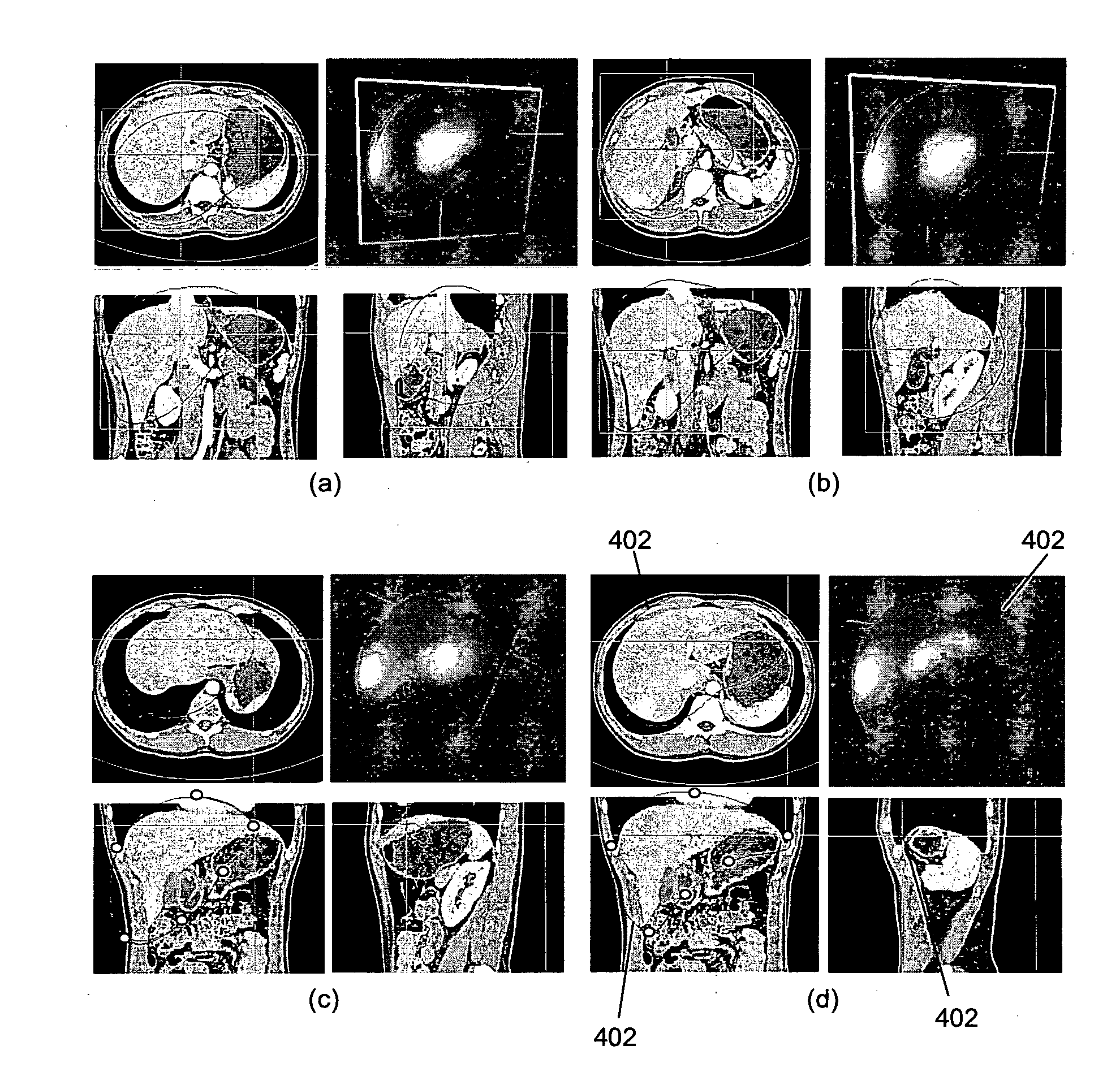
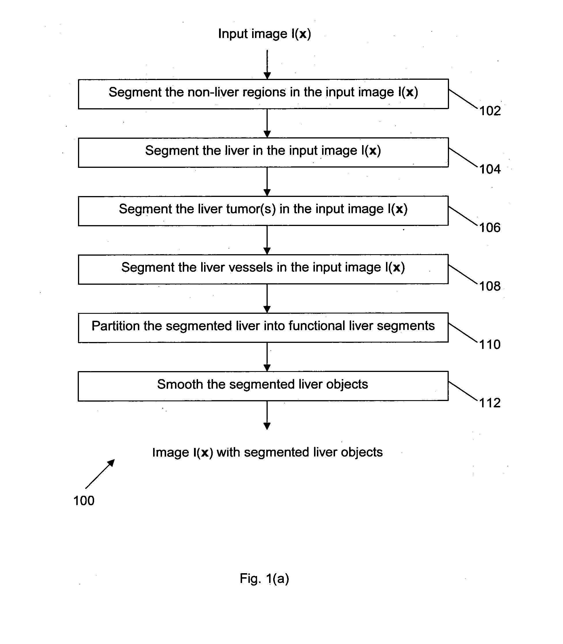
[0027]Referring to FIG. 1(a), the steps are illustrated of a method 100 which is an embodiment of the present invention, and which segments liver objects in an image.

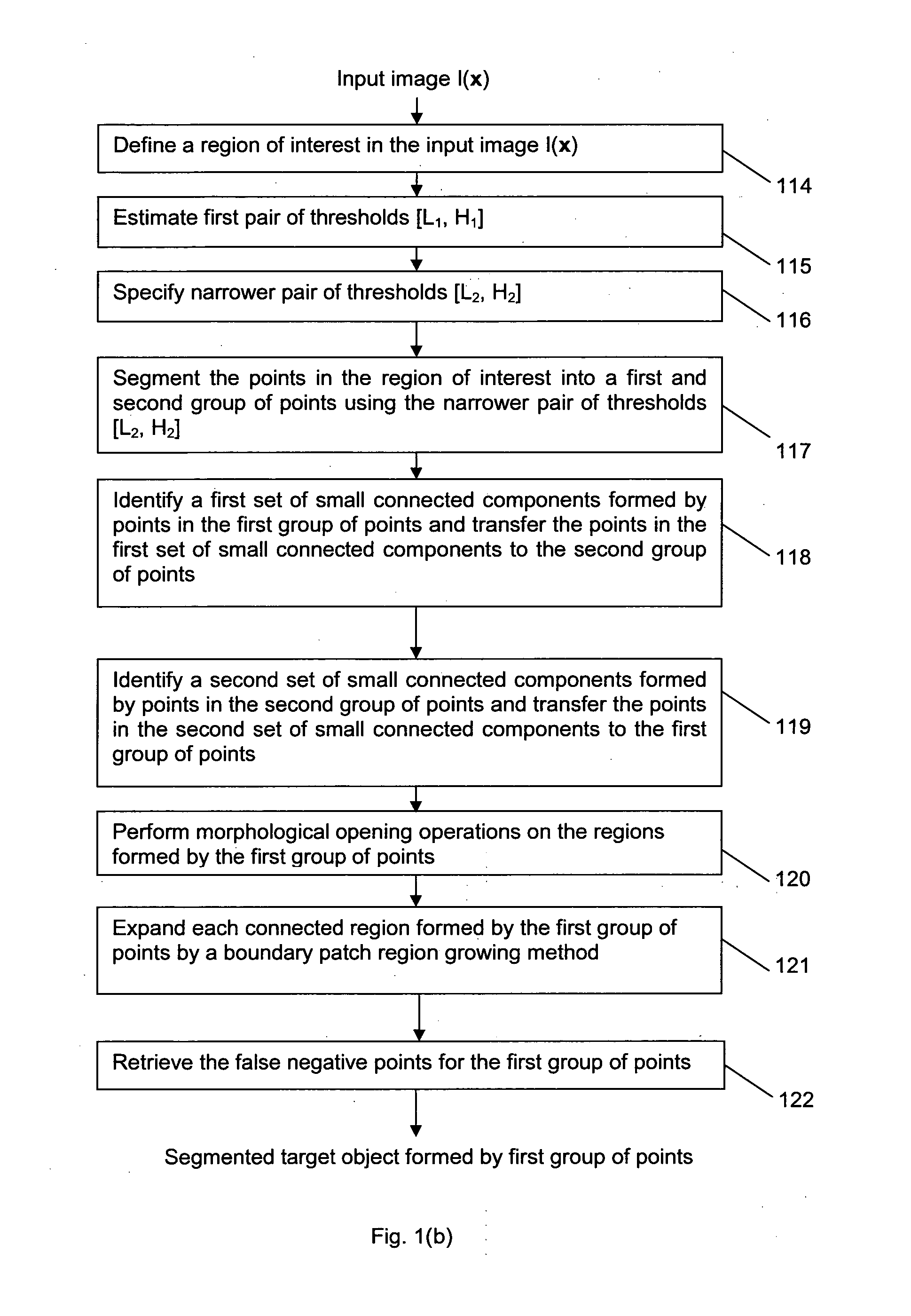
[0028]The input to method 100 is a three-dimensional image I(x) which may be a Computed Tomography (CT) image or any other type of image. The input image is defined in a domain D ⊂ R3 and comprises a plurality of points (voxels) having respective intensity values representing medical data. In step 102, the non-liver regions in the image I(x) are segmented. The sub-steps of step 102 are illustrated in FIG. 1(b). In step 104, the liver in the input image I(x) is segmented whereas in step 106, the liver tumor in the image I(x) is segmented (this may include segmenting multiple tumors if such tumors are present in the image). In step 108, liver vessels, in the image I(x) are segmented. In step 110, the segmented liver from step 104 is partitioned into functional liver segments and in step 112, the segmented liver objects (i...


PUM


Abstract

A method for segmenting an object of interest (which may be a liver object) in an input image is disclosed. The method comprises the steps of: (a) segmenting the points in a region of interest in the input image into a first and second group of points using predetermined intensity threshold values; (b) identifying a first set of small connected components formed by points in the first group of points and transferring the points in the first set of small connected components to the second group of points; (c) identifying a second set of small connected components formed by points in the second group of points and transferring the points in the second set of small connected components to the first group of points; and (d) forming the segmented object of interest using the first group of points.

Description

FIELD OF THE INVENTION [0001]The present invention relates to a method and system for segmenting objects in an image. The objects may be anatomy structures. The particular application example is the segmentation of liver objects in medical images, such as CT images.BACKGROUND OF THE INVENTION[0002]To achieve better results in the diagnosis, treatment and monitoring of liver diseases, such as liver failure due to cirrhosis and liver cancer (both primary and secondary), an accurate segmentation of liver structures (or liver objects) in medical images, such as CT images, is required. The liver objects may be for example, the liver itself, a tumor in the liver, vasculature structure in the liver (i.e. liver vessels) or functional segments of the liver.[0003]Manual segmentation of liver structures in CT images is not only time consuming, it also involves large inter- and intra-expert variations. Although automatic segmentation methods may be adopted instead, these automatic segmentation ...

Claims


Application Information

Patent Timeline
Patent Type & Authority Applications(United States)
IPC IPC(8): G06K9/00G06K9/34
CPCG06T7/0081G06T2207/10081G06T2207/30056G06T2207/20148G06T2207/20141G06T7/11G06T7/136G06T7/187
Inventor LIU, JIMIN
Owner AGENCY FOR SCI TECH & RES
Why Eureka
Try Eureka