
Antigen Binding Proteins

a technology of binding proteins and antigens, applied in the field of antigen binding proteins, can solve the problems of difficult separation from the desired heterodimer monobodies, formation of undesired homodimers,

Active Publication Date: 2012-09-20
F HOFFMANN LA ROCHE INC
View PDF6 Cites 29 Cited by
  • Summary
  • Abstract
  • Description
  • Claims
  • Application Information

AI Technical Summary

Benefits of technology

[0072]It has now been found that the antigen binding proteins according to the invention have valuable characteristics such as biological or pharmacological activities (as e.g. enhanced ADCC compared to parent antibodies). They can be used e.g. for the treatment of diseases such as cancer. The antigen binding proteins according to the invention have furthermore highly valuable pharmacokinetic properties (like e.g. AUC0-inf, Cmax or C0).

Problems solved by technology

However during expression a considerably amount of undesired homodimer is formed as by-product, which is difficult to separate from the desired heterodimeric monobodies, as the homodimer and the heterodimer have the same or similar molecular weights.

Method used


Image

  • Antigen Binding Proteins
  • Antigen Binding Proteins
  • Antigen Binding Proteins

Examples


example 1

Generation of MoAb-Dimer Antigen Binding Proteins

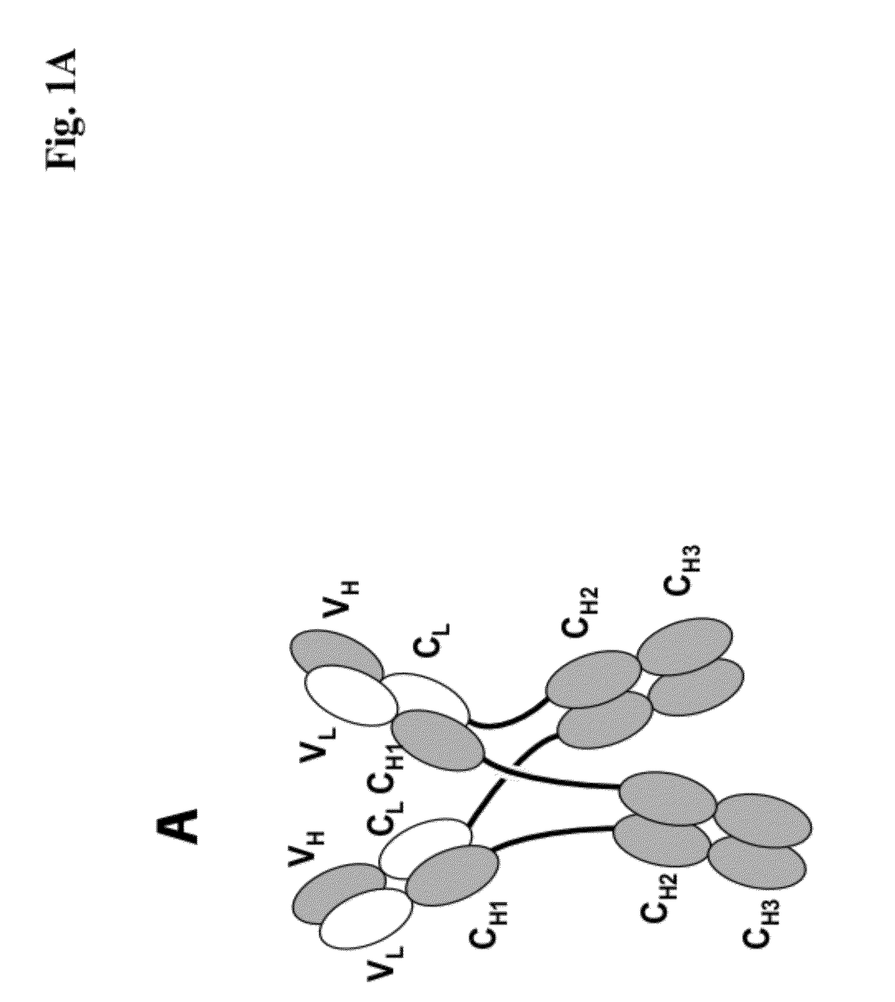
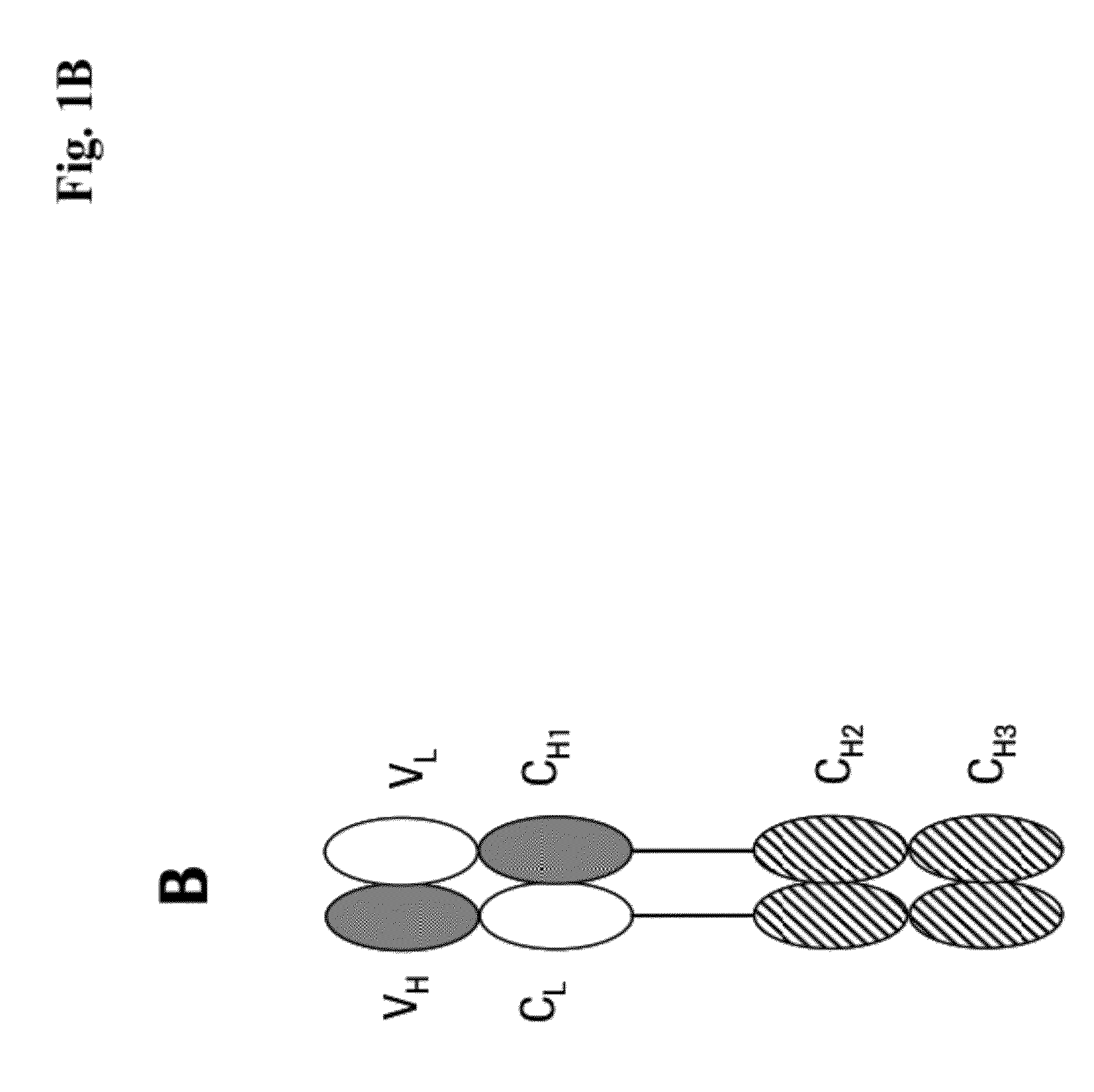
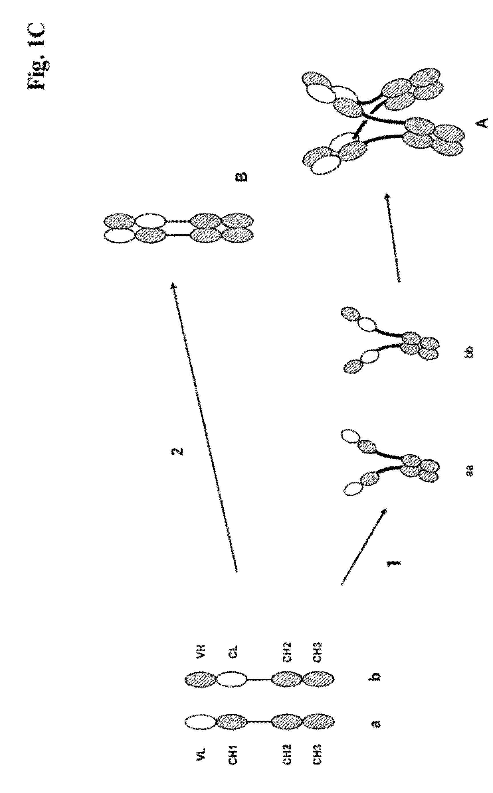
[0273]We designed antigen binding proteins according to the invention against c-Met (SEQ ID NO:1 and SEQ ID NO:2), IGF-1R (SEQ ID NO:3 and SEQ ID NO:4) and HER3 (SEQ ID NO:5 and SEQ ID NO:6) based on the design principle as shown in FIG. 1A. The respective constructs were transiently expressed in HEK293 cells as described above, and subsequently purified via Protein A affinity chromatography followed by size exclusion. FIGS. 2-4 depict the chromatograms of the size exclusion chromatography of the three different productions of antigen binding proteins as well as the corresponding SDS-PAGE under non-reducing and reducing conditions. In addition to peak 3 in FIG. 2, peak 2 in FIG. 3, peak 3 in FIG. 4 proteins eluting at earlier timepoints from the column peak 2 in FIG. 2, peak 1 in FIG. 3, peak 4 in FIG. 7 were observed. Based on their retention time it was calculated that they exhibited the double the molecular weight of the monovalent...

example 2

IGF-1R Binding Affinity

[0277]IGF-1R extracellular domain binding of the IGF1R AK18 MoAb-Dimer (“CH3-wt”) was compared to the binding of the parental IgG1 antibody by surface Plasmon resonance (SPR). FIG. 5 depicts the scheme of the SPR assay to determine the affinity. The analysis (double determination) showed that the IGF-1R binding affinity is retained in the IGF1R AK18 MoAb-Dimer (“CH3-wt”).

k (on)k (off)KDMab (IGF-1R)1.74E+06 6.63E−033.80E−09MoAb-Dimer (IGF-1R) (1. deter.)1.5E+06 3.0E−032.01E−09MoAb-Dimer (IGF-1R) (2. deter.)1.5E+06 3.0E−032.05E−09

example 3

Cellular Binding to IGF-1R Expressing Cell Lines

[0278]Cellular binding of IGF-1R MoAb-Dimer was demonstrated on A549 cells. A549 cells in the logarithmic growth phase were detached with accutase (Sigma) and 2×10e5 cells were used for each individual antibody incubation. IGF-1R antibody and MoAb-Dimer were added in a threefold dilution series (100-0.0003 μg / mL). Bound antibodies were visualized with a secondary Alexa488-coupled antibody (5 μg / mL) binding to the constant region of human immunoglobulin. Dead cells were stained with 7-AAD (BD) and excluded from the analysis. Fluorescence intensity of single cells was measured on a FACS Canto (BD Biosciences) flow cytometer. The data show that the MoAb-Dimer showed very similar halfmaximal binding to cells comparable to the parental IGF-1R IgG1 antibody. This implies that the MoAb-Dimer can bind with two arms to IGF-1R on cells and exhibits an avidity effect. However, the total mfi (mean fluorescence intensity) is higher for the MoAb-Dim...


PUM

PropertyMeasurementUnit
Fractionaaaaaaaaaa

Abstract

The present invention relates to antigen binding proteins comprising two Fc parts, methods for their production, pharmaceutical compositions containing said antigen binding proteins, and uses thereof.

Description

CROSS-REFERENCE TO RELATED APPLICATIONS[0001]The present application claims of priority under 35 USC §119(a) to European patent application number EP11156320.1, filed 28 Feb. 2011, the contents of which is incorporated herein by reference.SEQUENCE LISTING[0002]A sequence listing comprising SEQ ID NOS: 1-6 is attached hereto. Each sequence provided in the sequence listing is incorporated herein by reference, in its entirety, for all purposes.TECHNICAL FIELD[0003]The present invention relates to antigen binding proteins comprising two Fc parts, methods for their production, pharmaceutical compositions containing said antigen binding proteins, and uses thereof.BACKGROUND OF THE INVENTION[0004]In the last two decades various engineered antibody derivatives, either mono or- multispecific, either mono- or multivalent have been developed and evaluated (see e.g. Holliger, P., et al., Nature Biotech. 23 (2005) 1126-1136; Fischer, N., and Léger O., Pathobiology 74 (2007) 3-14).[0005]US 2004 / 0...

Claims


Application Information

Patent Timeline
IPC IPC(8): A61K39/395A61P35/00C12N5/10C12N15/13C12N15/85C07K16/18C12P21/00
CPCC07K16/00C07K16/2863C07K2317/64C07K2317/66C07K2317/732C07K2317/41C07K2317/77C07K2317/92C07K2317/94C07K16/32C07K2317/76A61P35/00G01N21/553C07K16/18A61K39/395C07K16/468C07K2317/72C12N15/85
Inventor BOSSENMAIER, BIRGITKETTENBERGER, HUBERTKLEIN, CHRISTIANKUENKELE, KLAUS-PETERREGULA, JOERG-THOMASSCHAEFER, WOLFGANGSCHWAIGER, MANFREDSUSTMANN, CLAUDIO
Owner F HOFFMANN LA ROCHE INC
Learn More