

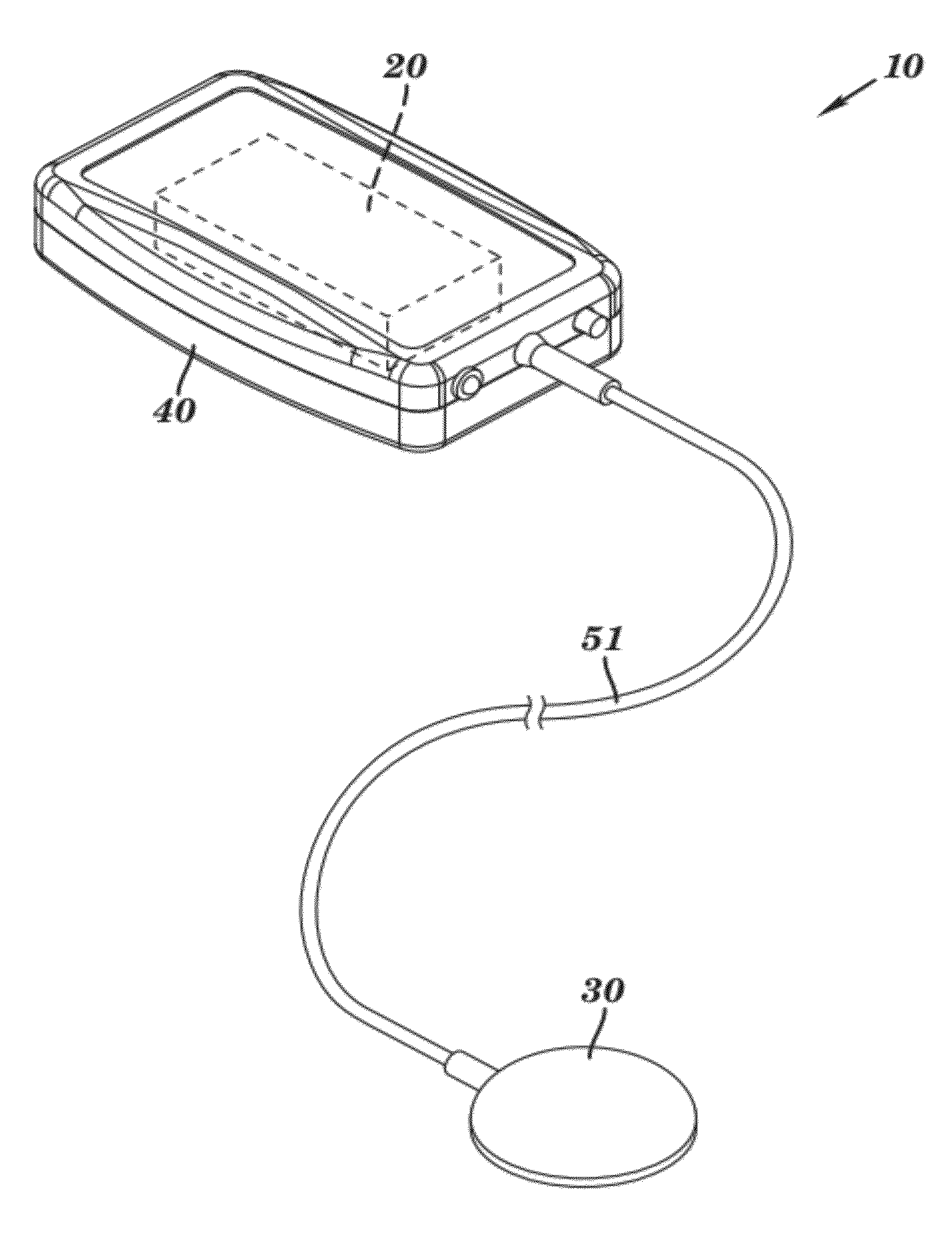
Portable ultrasound system
a portable ultrasound and ultrasound technology, applied in ultrasonic/sonic/infrasonic diagnostics, applications, etc., can solve the problems of inability to use for long periods of time, inability to carry and carry therapeutic ultrasound devices in the art, and inability to achieve long-term use of therapeutic ultrasound devices, so as to improve the overall quality of life, reduce recovery time, and increase flexibility
- Summary
- Abstract
- Description
- Claims
- Application Information
AI Technical Summary
Benefits of technology
Problems solved by technology
Method used
Image

Examples
example 1
Device Risk Assessment For Studying Pain Reduction Using Continuous Low Intensity Ultrasound
[0139]Ultrasound is currently used in many medical diagnostic applications across the globe such as imaging, fetal heart rate monitoring, and blood flow analysis. Ultrasound is also present in various non-diagnostic drug delivery and therapeutic applications. The mechanical and thermal mechanisms of action in ultrasound have been shown to facilitate wound and bone fracture healing, enhance the penetration of topical ointments into the skin, provide pain and healing relief in physiotherapy, and perform non-invasive tumor and fibroid ablation.
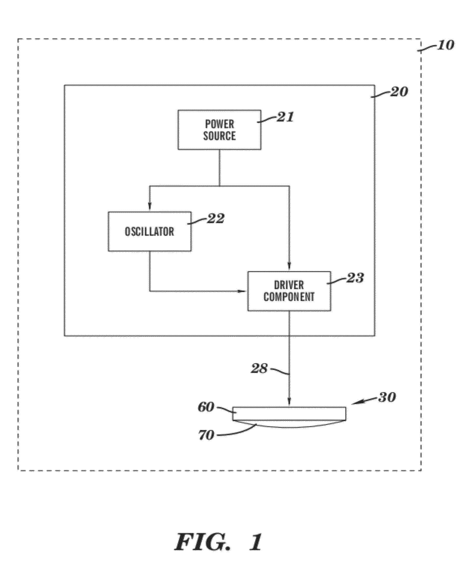
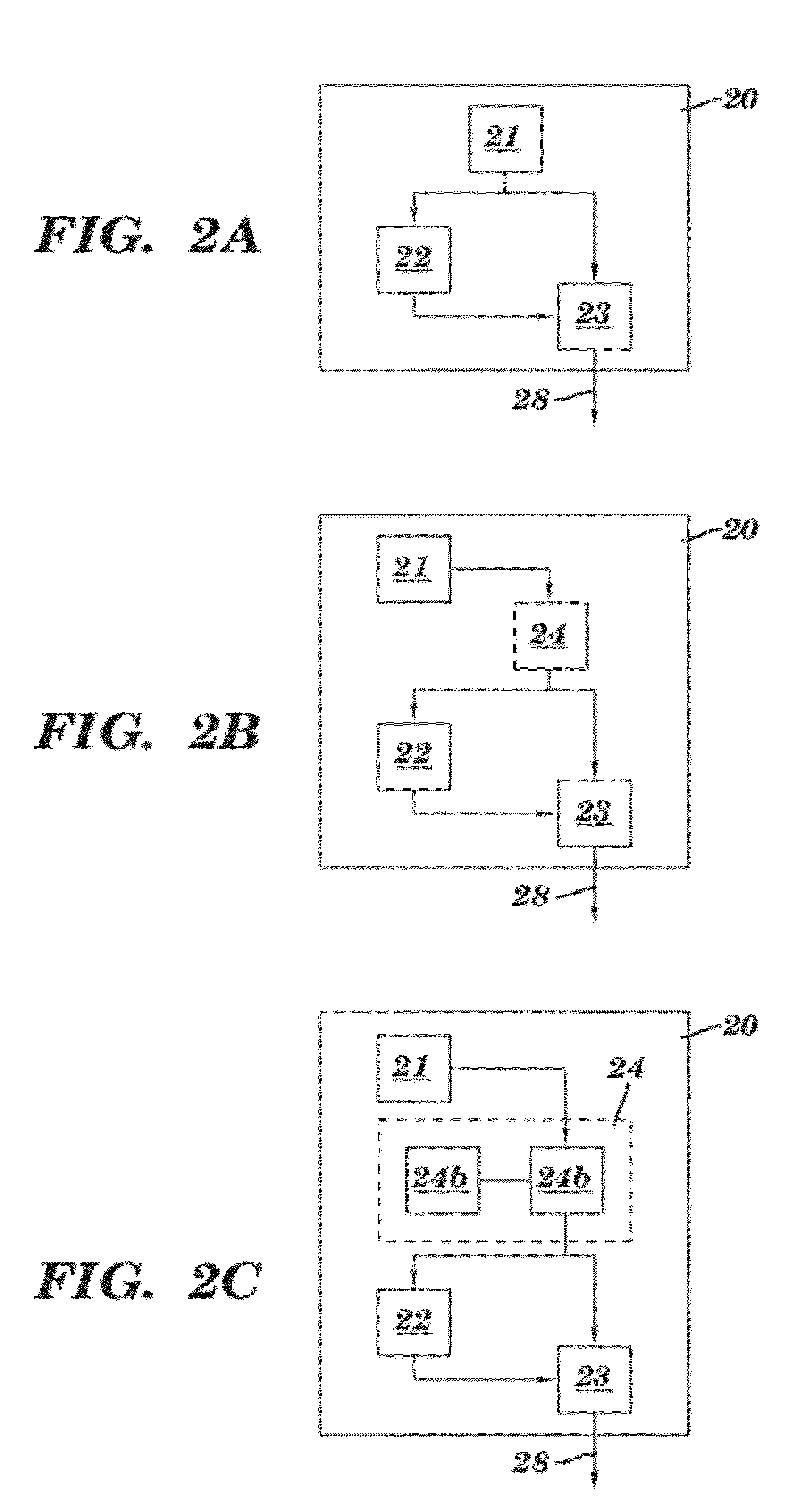
[0140]Aggressive miniaturization and full system integration of the ultrasound transducer, electronics, and power supply has provided a unique platform solution for using ultrasound in frontline medicine. TheraSonX™ is the first truly portable Low Intensity Ultrasound (LIUS) device which can provide safe, effective relief outside of the hospital. The risk ...
example 2
UltrOZ Therapeutic Case Study: Splint Reduction
[0243]History: Subject was a 2 yr old thoroughbred filly. The filly had been in race training on a farm and was moving up to the next level of training by coming to the Churchill Downs race track.
[0244]Evaluation: The filly arrived at the track with a large splint on the right front leg, medial aspect. The filly was not lame on arrival or through the days of UltrOZ ultrasound treatment.
[0245]Treatment: The UltrOZ unit was applied to the caudal aspect of the splint, covering the suspensory ligament as well. It was applied from 10 a.m. to 3 p.m. every day, 7 days a week from 10 / 22 to 11 / 20. No other treatments were performed.
[0246]Outcome: The filly never exhibited any soreness in the suspensory ligament, which is unusual with splints this size. The splint was reduced in size by 30% over the 4 week period of use. The trainer and Equine Therapist were very happy with the results of treatment. The reduction of splint size during an increase...
example 3
Horse Case Study
[0247]Horse Subject: 9 year old, thoroughbred, gelding. Mid level event horse.
[0248]History: Acute presentation of ⅗ lameness on the right forelimb.
[0249]Assessment: A new splint had formed on the proximal aspect of the 4th metacarpal bone of the right fore. The horse blocked to regional local anesthesia of the splint and suspensory ligament. Radiographs revealed no fracture but active periosteal proliferation of the splint. Ultrasound of the suspensory ligament showed mild inflammation at the region associated with the splint.
[0250]Treatment: The horse was treated with the anti-inflammatory Equioxx for 10 days. A regional injection at the splint / suspensory interface of betamethasone / serapin / predef was done. The horse was stall rested for 7 days, stalled and tack walked for 7 days. Small paddock with tack walk for remainder of 30 days.
[0251]A prototype Zetroz therapeutic ultrasound unit was utilized beginning 2 days after the initial injection for 3 weeks. The unit w...
PUM
| Property | Measurement | Unit |
|---|---|---|
| angle | aaaaa | aaaaa |
| voltages | aaaaa | aaaaa |
| frequencies | aaaaa | aaaaa |
Abstract
Description
Claims
Application Information