
Method and device for adjusting the visualization of volume data of an object

Active Publication Date: 2012-12-06
SIEMENS HEALTHCARE GMBH
View PDF22 Cites 7 Cited by
  • Summary
  • Abstract
  • Description
  • Claims
  • Application Information

AI Technical Summary

Benefits of technology

[0017]The present embodiments may obviate one or more of the drawbacks or limitations in the related art. For example, th

Problems solved by technology

Despite this combination of projections taken from different angles, conventional mammography has its limits.
There is the risk that tissue hardening (e.g., calcifications) is co

Method used


Image

  • Method and device for adjusting the visualization of volume data of an object
  • Method and device for adjusting the visualization of volume data of an object
  • Method and device for adjusting the visualization of volume data of an object

Examples


Example

DETAILED DESCRIPTION OF THE DRAWINGS

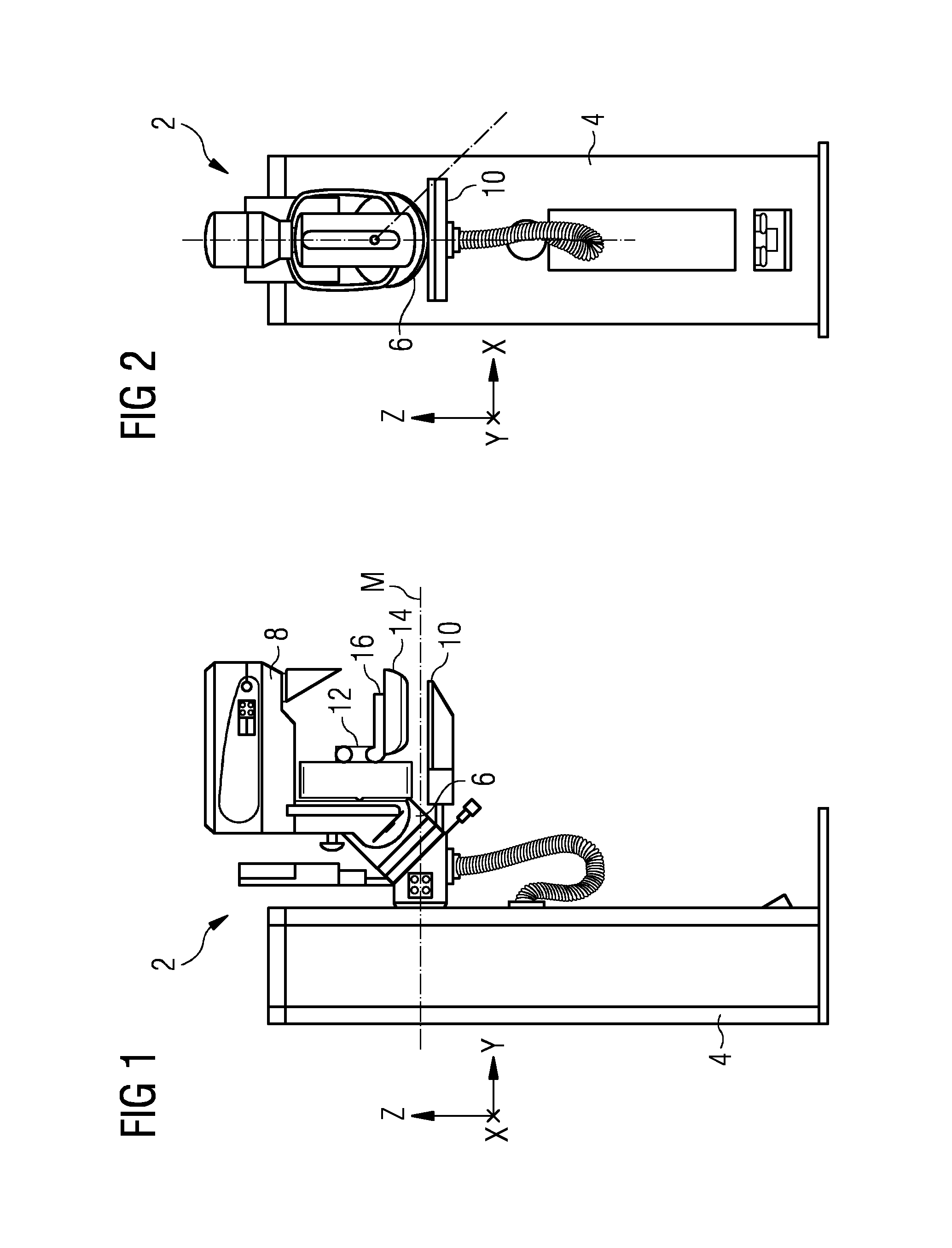
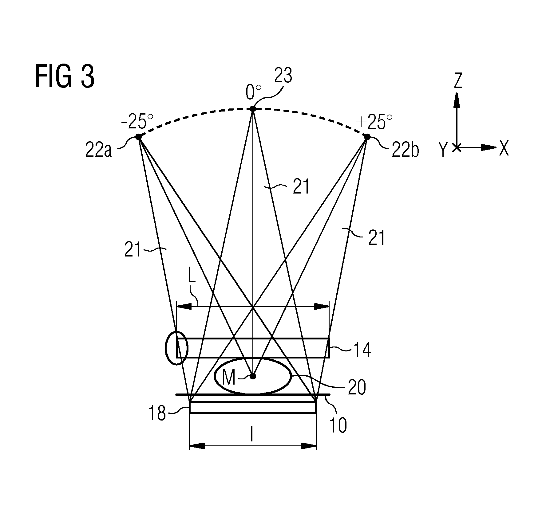
[0029]FIGS. 1 and 2 show a side view and a front view, respectively, of a mammography device 2. The mammography device 2 has a base body embodied as a stand 4 and, projecting out from the stand 4, an angled device arm 6. An irradiation unit 8 embodied as an X-ray emitter is arranged at a free end of the angled device arm 6. Also mounted on the device arm 6 are an object table 10 and a compression unit 12. The compression unit 12 includes a compression element 14 that is arranged relative to the object table 10 and is displaceable along a vertical Z-direction. The compression unit 12 also includes a support 16 for the compression element 14. In this arrangement, a type of lift guide is provided in the compression unit 12 for the purpose of moving the support 16 together with the compression element 14. Additionally, arranged in a lower section of the object table 10 is a detector 18 (cf. FIG. 3) that in the present exemplary embodiment is a digital...


PUM


Abstract

An adjustment of the visualization of volume data of an object as an image with regard to diagnostically relevant medical information relating to the environment of a region under investigation is provided. In the process, at least one slice region is specified in accordance with slice information within the volume data. A first mapping of a value range of the volume data is used for visualizing the at least one slice region on a display. A second mapping different from the first mapping is used for visualizing a region bordering on the at least one slice region on the display.

Description

[0001]This application claims the benefit of DE 10 2011 076 930.7, filed on Jun. 3, 2011.BACKGROUND[0002]The present embodiments relate to a method, a device and a computer program for adjusting the visualization of volume data of an object as an image.[0003]The use of X-rays in medical diagnosis is a widely established practice. Thus, for example, the examination of female breast tissue for the formation of carcinomas may be carried out using X-ray radiation (e.g., mammography).[0004]Owing to the specific anatomical features of the region of the body being examined, special-purpose devices, which may be referred to as mammography devices, are used for examinations of the type using X-rays.[0005]Projection settings of mammography devices have become established as standard settings for diagnostic purposes. The following two standard settings may be used.[0006]The mediolateral oblique (MLO) view of the breast (e.g., oblique projection) is the standard setting employed in the early de...

Claims


Application Information

Patent Timeline
IPC IPC(8): G06K9/36
CPCA61B6/025A61B6/03A61B6/037A61B6/4441A61B6/463A61B6/466G06T15/08A61B6/502A61B6/5217A61B6/5223A61B8/0825A61B8/13A61B8/463A61B6/469
Inventor ENGEL, KLAUSJEREBKO, ANNA
Owner SIEMENS HEALTHCARE GMBH
Learn More