AI technical title is built by Patsnap AI team. It summarizes the technical point description of the patent document.
a technology of helix tissue and grasping device, which is applied in the direction of osteosynthesis device, surgical staple, surgical forcep, etc., can solve the problems of difficult use in endoscopic procedures, and achieve the effect of better visualization
Active Publication Date: 2013-01-03
APOLLO ENDOSURGERY INC
View PDF1 Cites 8 Cited by
Summary
Abstract
Description
Claims
Application Information
AI Technical Summary
This helps you quickly interpret patents by identifying the three key elements:
Problems solved by technology
Method used
Benefits of technology
Benefits of technology
[0017]In accordance with another aspect of the present invention there is provided an endoscopic suturing system for use with an endoscope having a cap assembly adapted to be positioned at the distal end of the endoscope where the cap assembly includes an elongate needle guard. The needle guard generally extends from a base of the cap in a direction distal to the end of the endoscope. Preferably the needle guard extends in a distal direction parallel to the axis of the endoscope. The needle guard is adapted to prevent tissue from inadvertently contacting the needle tip while the needle tip is in an open position and the tissue is being positioned for suturing.
[0018]In accordance with another aspect of the present invention there is provided an endoscopic suturing system for use with an endoscope having a cap assembly adapted to be positioned at the distal end of the endoscope where the cap assembly includes an elongate channel guard. The channel guard generally extends from a base of the cap in a direction distal to the end of the endoscope and is coaxial with the endoscope channel which used by the needle capture device. The channel guard is adapted to aid in suturing by positioning tissue a sufficient distance away from the end of the endoscope channel allowing for better visualization and providing a surface to support the tissue during the suturing operation. Preferably, the distal end of the channel guard is inclined to provide a plane which is generally perpendicular to the needle tip as the needle tip intersects the plane along the needle suturing path. Preferably, the minimum length that the channel guard extends from the cap is related to the field of view from the endoscope such that minimum length allows sufficient tissue to be visualized when the tissue is placed in a position for suturing.
Problems solved by technology
While this system affords the ability to grasp thick tissue, the tissue grasping arm and the arrangement of the needle recovery member provides bulk to the system making it difficult to use in endoscopic procedures.
Method used
Image
Click on the blue label to locate the original text in one second.
Reading with bidirectional positioning of images and text.
Examples
Embodiment Construction
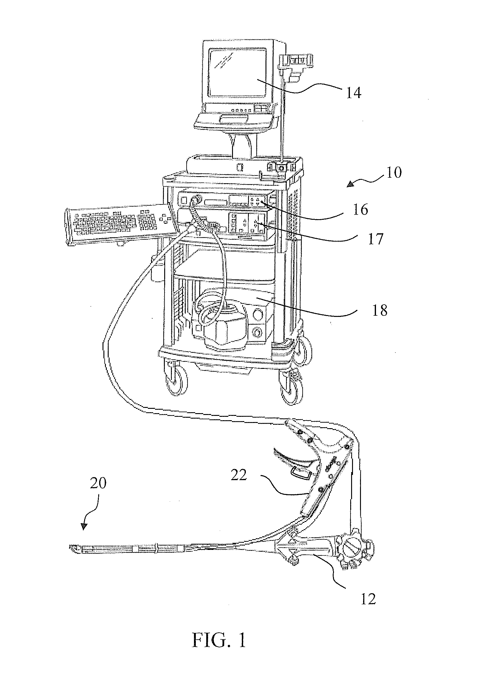
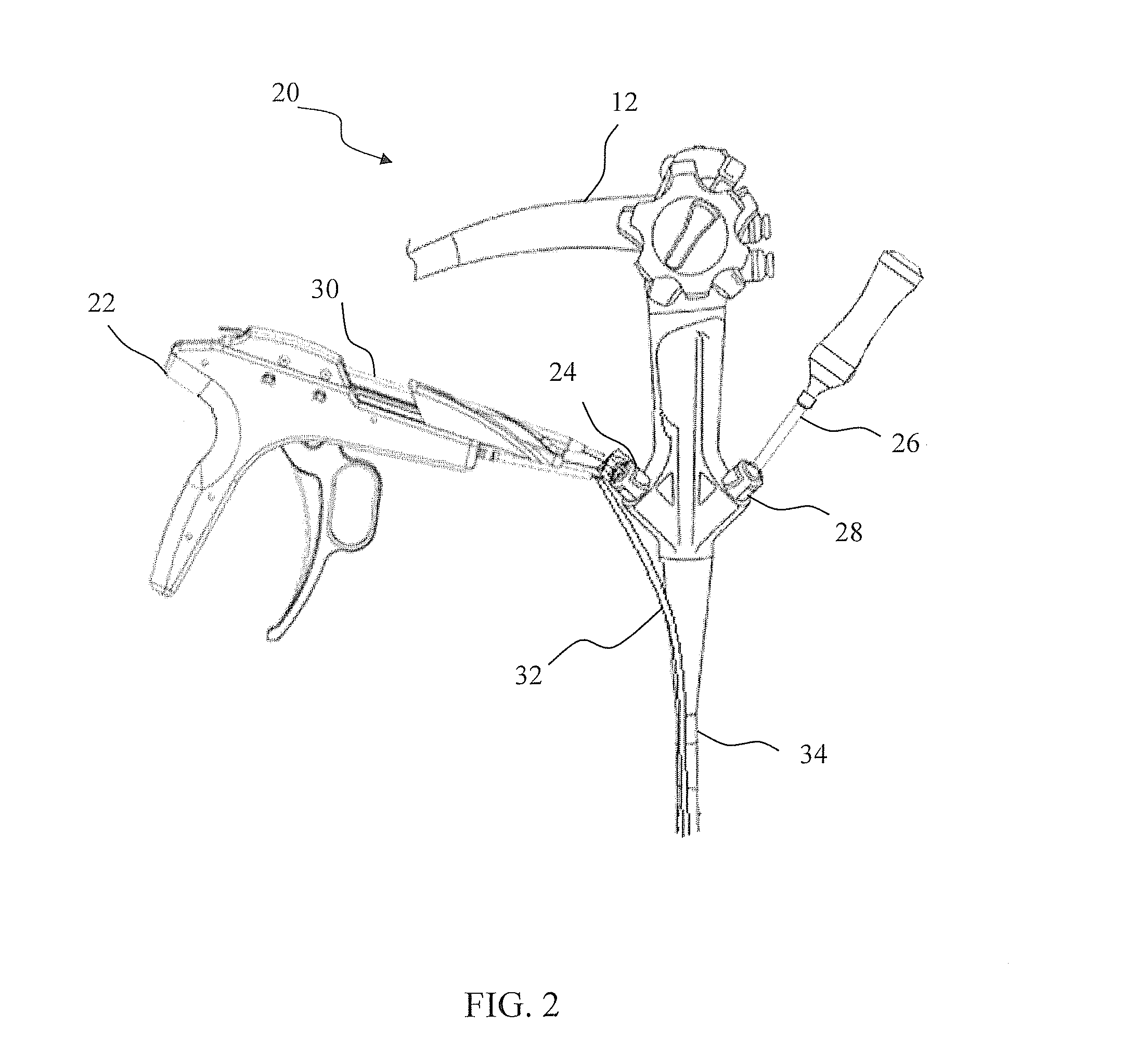
[0197]As shown in FIG. 1 an endoscope system 10 which comprises an endoscope 12, a video display unit 14, an image processing device 16, a light source 17, a suction device 18 is used with and an endoscopic suturing device 20 as part of an endoscopic treatment system according to one embodiment of the present invention. FIG. 2 and FIG. 3 illustrate respectively the proximal and distal portions of endoscope 12 and endoscopic suturing device 20. The endoscopic suturing device 20 has an operable handle 22 which is removably coupled to endoscope 12 at a first instrument channel 24. A tissue grasper 26 which is used to gather tissue is shown positioned within a second instrument channel 28 of endoscope 12. The endoscopic suturing device 20 includes an elongate needle capture device 30 which is removably coupled to handle 22 and extends to the distal end of endoscope 12 slidably positioned within instrument channel 24. The endoscopic suturing device 20 is operated by handle 22 which is pr...
PUM
Abstract
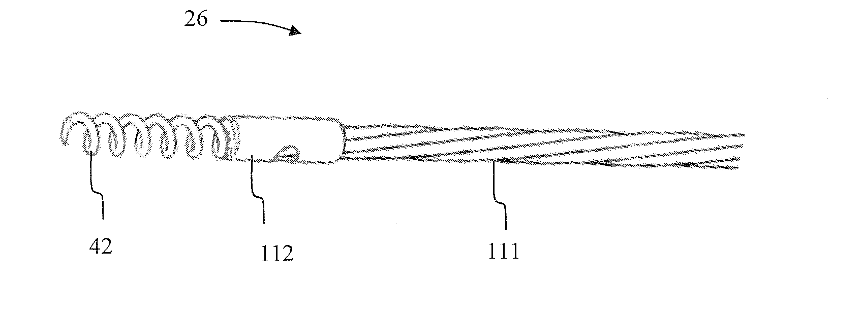
An endoscopic tissue grasper device includes a flexible tubular member, a flexible shaft extending through the tubular member, a proximal handle for moving the shaft and tubular member relative to each other, and a distal helical coil having a sharpened end for engaging tissue. The helical coil has a proximal close wound portion, a distal open wound portion, and an intermediate transition portion between the proximal and distal portions. The flexible shaft extends into the proximal and intermediate portions and is secured thereto, which prevents tissue engaged by the helical coil from becoming wedged in the transition portion and thereby facilitates release of the coil from engaged tissue. In addition, a bearing sheath is provided between the shaft and tubular member to prevent the shaft from binding during operation.
Description
CROSS-REFERENCE TO RELATED APPLICATIONS[0001]This application is a continuation of U.S. Ser. No. 13 / 539,661, filed Jul. 2, 2012, which is a continuation-in-part of U.S. Ser. No. 13 / 328,003 filed Dec. 16, 2011, which claims priority from U.S. Prov. Ser. No. 61 / 483,679 filed May 8, 2011 and U.S. Prov. Ser. No. 61 / 495,970 filed Jun. 11, 2011, which is a continuation-in-part of U.S. Ser. No. 12 / 485,576 filed Jun. 16, 2009, which claims priority from U.S. Prov. Ser. No. 61 / 073,340 filed Jun. 17, 2008 and U.S. Prov. Ser. No. 61 / 162,249 filed Mar. 20, 2009, all of which are hereby incorporated by reference herein in their entireties.BACKGROUND OF THE INVENTION[0002]1. Field of the Invention[0003]The present invention relates to a treatment device which can be inserted into a body through a natural orifice with an endoscope or other steerable guide member. The present invention may be used to perform suturing on the tissue of a mammal, whether human or not, and whether or not alive, but is ...
Claims
Application Information
Patent Timeline
Application Date:The date an application was filed.
Publication Date:The date a patent or application was officially published.
First Publication Date:The earliest publication date of a patent with the same application number.
Issue Date:Publication date of the patent grant document.
PCT Entry Date:The Entry date of PCT National Phase.
Estimated Expiry Date:The statutory expiry date of a patent right according to the Patent Law, and it is the longest term of protection that the patent right can achieve without the termination of the patent right due to other reasons(Term extension factor has been taken into account ).
Invalid Date:Actual expiry date is based on effective date or publication date of legal transaction data of invalid patent.
Patent Type & AuthorityApplications(United States)

