
2d-2d fusion for interventional guidance in trans-catheter aortic valve implantation

a technology of interventional guidance and fusion, which is applied in the field of 2d2d fusion for interventional guidance in transcatheter aortic valve implantation, can solve the problems of difficult 3d model to 2d fluoroscopy, may not always be available in all tavi cases, etc., and achieve the effect of maximizing the similarity measur

Active Publication Date: 2013-03-07
SIEMENS HEALTHCARE GMBH
View PDF6 Cites 13 Cited by
  • Summary
  • Abstract
  • Description
  • Claims
  • Application Information

AI Technical Summary

Benefits of technology

The invention is a method for improving the accuracy of detecting the shape of an anatomical structure in a medical image. The method involves creating a predefined shape model of the structure and then estimating the size of the structure in the image. The method then suggests a ratio between the size of the model and the actual size of the structure in the image to optimize the accuracy of the detection. The result is a more accurate and reliable way to detect the shape of anatomical structures in medical images.

Problems solved by technology

Although recent advances in utilizing three dimensional (3D) models to provide anatomical details further facilitate TAVI, 3D models may not always be available in all TAVI cases.
In addition, accurate registration of the 3D model to the 2D fluoroscopy presents a challenge.

Method used


Image

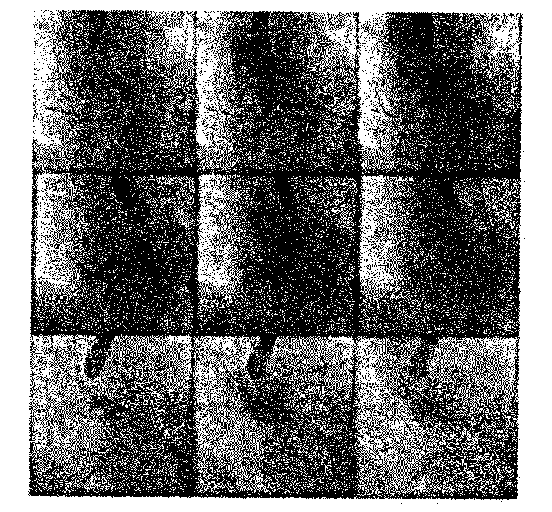
  • 2d-2d fusion for interventional guidance in trans-catheter aortic valve implantation
  • 2d-2d fusion for interventional guidance in trans-catheter aortic valve implantation
  • 2d-2d fusion for interventional guidance in trans-catheter aortic valve implantation

Examples


Embodiment Construction

[0023]Exemplary embodiments of the invention as described herein generally include systems and methods for 2D-2D fusion of a fluoroscopic frame without contrast injection with an angiographic frame showing the aortic root during TAVI procedures. Accordingly, while the invention is susceptible to various modifications and alternative forms, specific embodiments thereof are shown by way of example in the drawings and will herein be described in detail. It should be understood, however, that there is no intent to limit the invention to the particular forms disclosed, but on the contrary, the invention is to cover all modifications, equivalents, and alternatives falling within the spirit and scope of the invention.

[0024]As used herein, the term “image” refers to multi-dimensional data composed of discrete image elements (e.g., pixels for 2-dimensional images and voxels for 3-dimensional images). The image may be, for example, a medical image of a subject collected by computer tomography...


PUM


Abstract

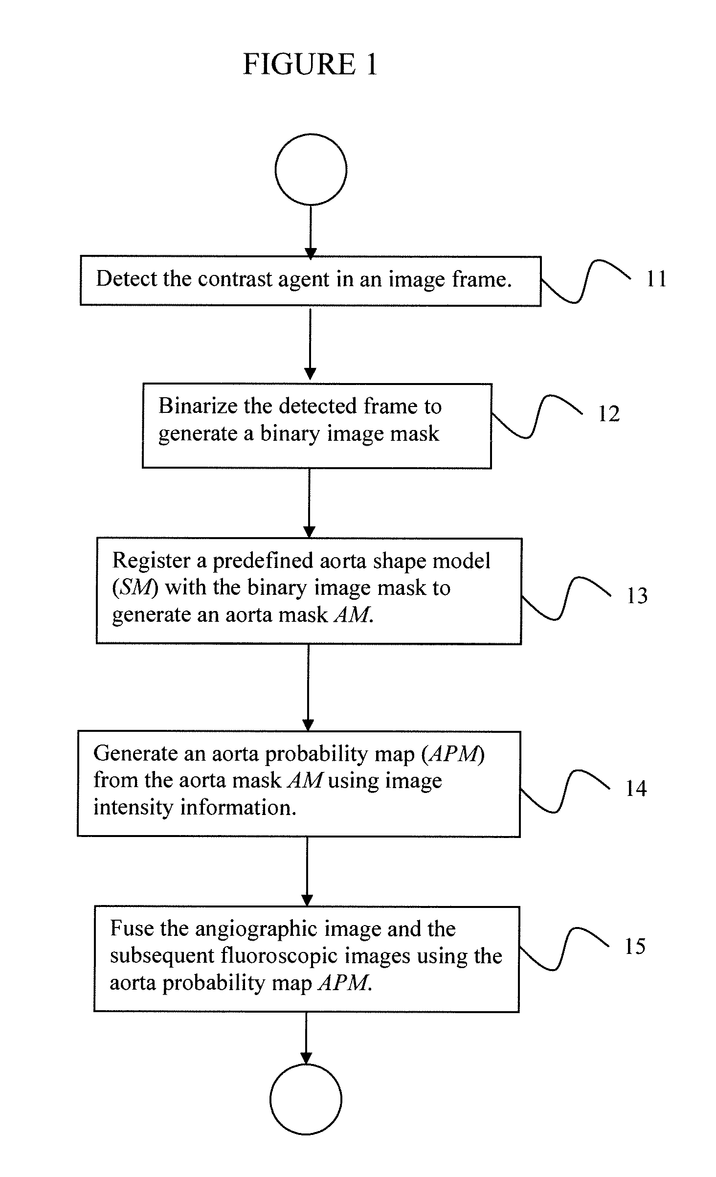
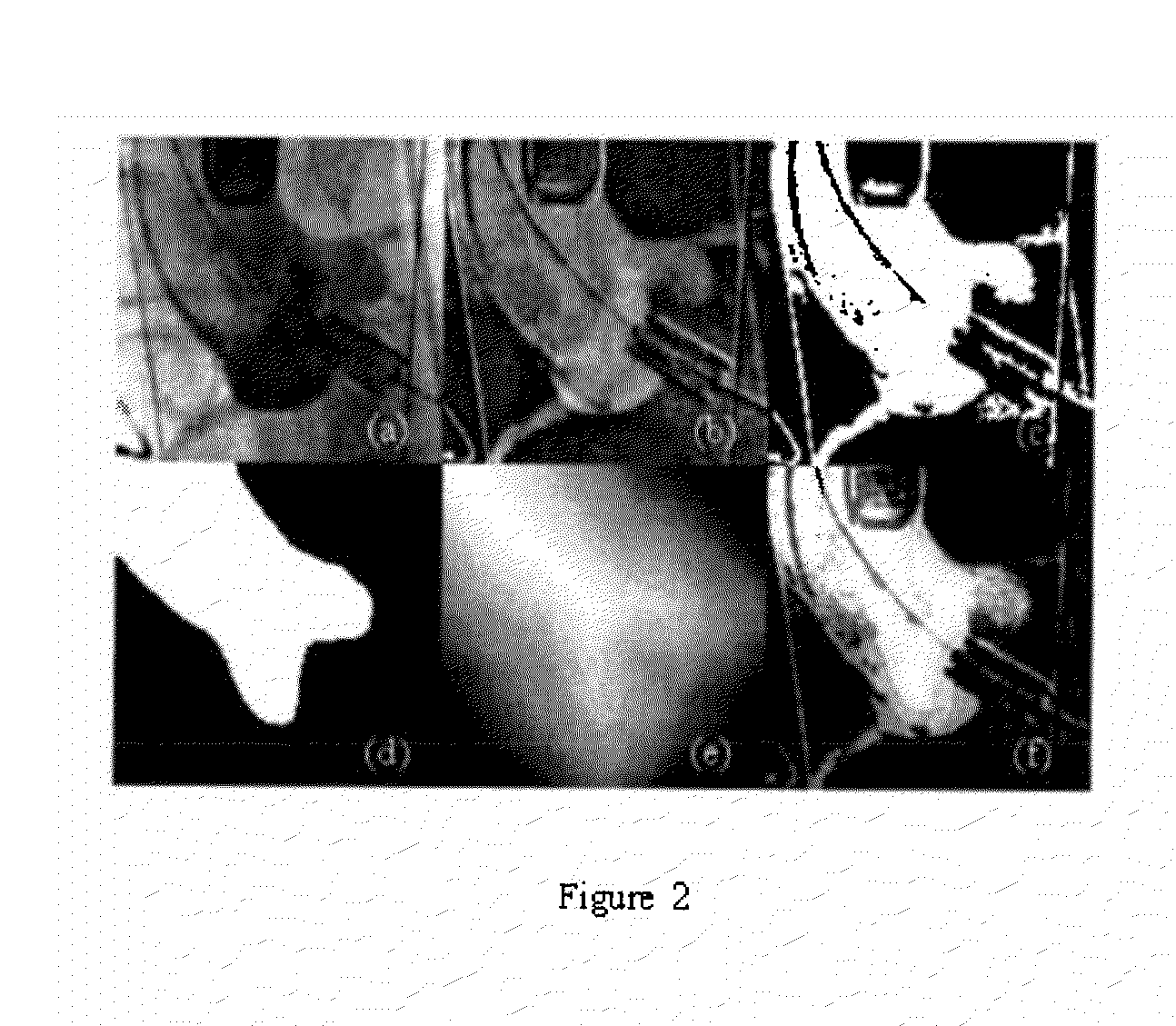
A method for fusing 2D fluoroscopic images with 2D angiographic images for real-time interventional procedure guidance includes using a 2-dimensional angiographic image to detect injection of a contrast agent into an anatomical structure, where the contrast agent first appears in a detected image frame, subtracting a background image from the detected image frame and binarizing the subtracted image frame to segment the anatomical structure from the subtracted image frame, registering the segmented anatomical structure with a predefined shape model of the anatomical structure to generate an anatomical mask, generating an anatomical probability map from the anatomical mask using intensity information from the subtracted image frame, where the anatomical probability map expresses a probability of a pixel in the subtracted image frame belonging to the anatomical structure, and fusing the angiographic image with one or more subsequently acquired 2-dimensional fluoroscopic images of the anatomical structure using the anatomical probability map.

Description

CROSS REFERENCE TO RELATED UNITED STATES APPLICATIONS[0001]This application claims priority from “2D2D Fusion for Interventional Guidance in TAVI Based on Contrast Detection and Aortic Root Localization”, U.S. Provisional Application No. 61 / 531,722 of Liao, et al., filed Sep. 7, 2011, the contents of which are herein incorporated by reference in their entirety.TECHNICAL FIELD[0002]This disclosure is directed to methods for visualizing target anatomy for real-time guidance of medical procedures.DISCUSSION OF THE RELATED ART[0003]Aortic valve disease affect about 1.8% of the global population, and is the most frequent heart valve disease in developed countries, leading to 60,000 surgical aortic valve replacements every year in Europe and even more in the United States. Trans-catheter aortic valve implantation (TAVI) is a new and breakthrough minimally invasive alternative to open heart surgery in patients with severe aortic stenosis. Two dimensional (2D) fluoroscopy is the typical ima...

Claims


Application Information

Patent Timeline
Patent Type & Authority Applications(United States)
IPC IPC(8): G06K9/36G09G5/02G06V10/26
CPCG06T3/0068G06T2207/30104G06K9/6289G06K9/6215A61B6/12A61B6/463A61B6/481A61B6/487A61B6/503A61B6/504A61B6/5235G06T5/50G06T2207/10121G06T2207/20076G06T2207/30048G06K9/34G06V10/26G06V10/761G06V10/803G06F18/251G06F18/22G06T3/14A61B6/00
Inventor LIAO, RUILIU, YINXIAOJOHN, MATTHIAS
Owner SIEMENS HEALTHCARE GMBH
Learn More