
Registration of thoracic and abdominal imaging modalities

a registration technology, applied in the field of thoracic and abdominal imaging registration, can solve the problems of not using a rigid approach with significantly more degrees of freedom, and achieve the effects of accurate registering, accurate registering, and accurate registering

Inactive Publication Date: 2008-07-08
SEGAMI R L
View PDF21 Cites 49 Cited by
  • Summary
  • Abstract
  • Description
  • Claims
  • Application Information

AI Technical Summary

Benefits of technology

[0035]T˘=arg⁢⁢max⁢⁢ST⁢(I,T⁡(J))Where I, J are the two images or volumes to register, and {hacek over (T)} is the transformation which maximizes an appropriately chosen similarity measure between images to register in a defined transformation space, with S the similarity function that provides this measure.
[0050]A final objective of this invention is to provide a method for accurate registering of anatomical and functional images which is rapid, requires minimal computational facilities, and is inexpensive to operate.

Problems solved by technology

However, in thoracic and abdominal applications, due to local deformations, movement of the organs, and the high variability of acquisitions, a non-rigid approach with significantly more degrees of freedom is needed.

Method used


Image

  • Registration of thoracic and abdominal imaging modalities
  • Registration of thoracic and abdominal imaging modalities
  • Registration of thoracic and abdominal imaging modalities

Examples


example 1

[0096]A first set of 11 CT, emission and transmission PET scans of the thoracic and abdominal regions were analyzed using the method of this patent application. This was a inhomogeneous data set because the data were derived a three different sites and varying machines and clinical protocols were used. A second set of 4 images were obtained using a combination PET-CT acquisition system, the Discovery LS PET / CT System, obtained from General Electric Medical Systems, Waukesha Wis. The process used for analysis has been designed to be independent of the scan parameters. In general, the CT images had a resolution of 512×512 pixels in the xy plane and between 46 and 103 slices, with voxel dimensions approximately 0.74×0.74×7.5 mm3. PET images had a resolution of 128×128 pixels in the xy plane with 163 to 205 slices, with voxel dimensions around 4.297×4.297×4.297 mm2. In the PET-CT combined machine data set, the CT images had a resolution of 512×512 and the PET images 128×128 pixels in th...

example 2

[0101]A third set of 18 CT, emission and transmission PET scans of the thoracic region were analyzed using the method of this patent application. This was a inhomogeneous data set because the data were derived a three different sites and varying machines and clinical protocols were used. The process used for analysis has been designed to be independent of the scan parameters. In general, the CT images had a resolution of 256×256 pixels in the xy plane and between 60 and 125 slices, with voxel dimensions approximately 1.0×1.0×5.0 mm2. PET images had a resolution of 144×144 pixels in the xy plane with 160 to 230 slices, with voxel dimensions around 4.0×4.0×4.0 mm3.

[0102]The evaluation method of Example 1 was used with Example 2. Table 2 shows the performance of this evaluation method for the different anatomical regions.

[0103]

TABLE 2RegionMeanVarianceChest Wall0.6700.02Mediastinal Wall (heart)0.9350.09Diaphragmatic Wall (liver)0.7200.11

[0104]Table 2 shows that the evaluation method is...


PUM


Abstract

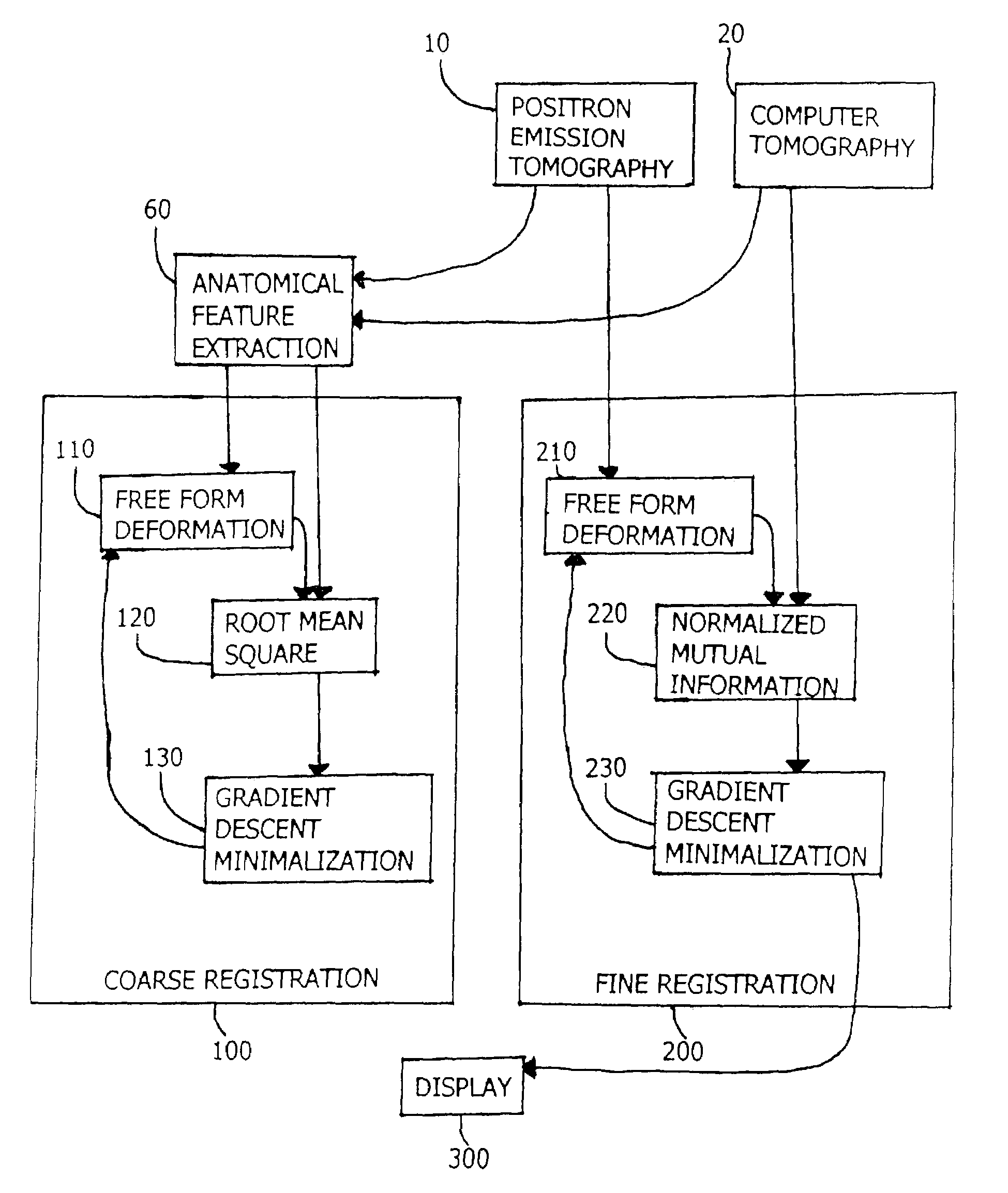
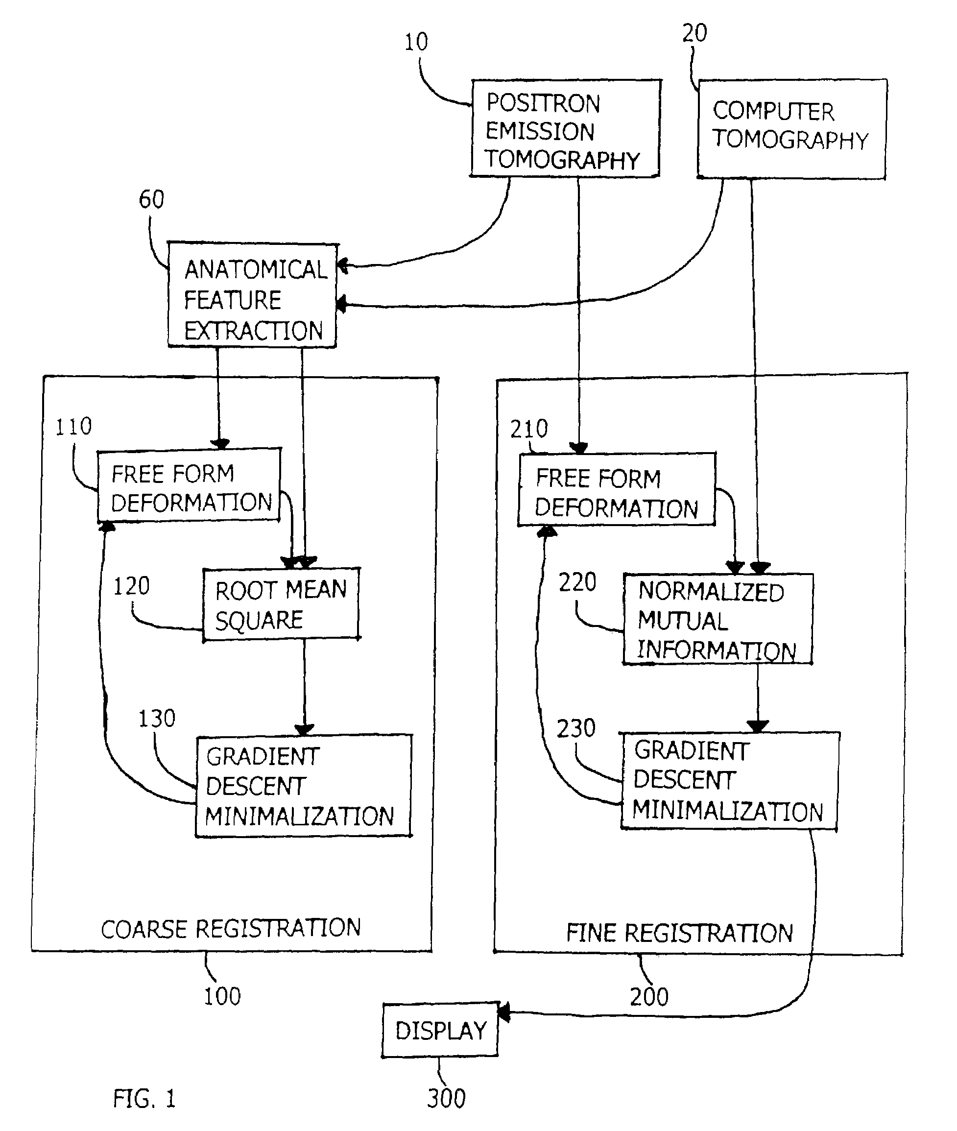
This disclosure presents an improved method for registering anatomical medical images and functional medical images. The example deals with the registration of x-ray computer tomography images with positron emission tomography images. The process is characterized by clinically useful registration with minimal computer calculations and minimal delay for computation. A nonrigid B-Spline free form deformation is used in both a preliminary coarse registration and the finished fine registration. Additional steps are used to insure accurate and complete registrations.

Description

CROSS-REFERENCE TO RELATED APPLICATIONS[0001]This application claims priority from U.S. provisional patent application Ser. No. 60 / 369,681 filed Apr. 3, 2002.STATEMENT REGARDING FEDERALLY SPONSORED RESEARCH OR DEVELOPMENT.[0002]Not Applicable.Reference to a “Microfiche Appendix.”[0003]Not Applicable.BACKGROUND OF THE INVENTION[0004]1. Field of the Invention[0005]This invention relates to methods for correlating clinical thoracic and abdominal images obtained by positron emission tomography (PET) and computer tomography (CT).[0006]2. Description of Related Art Including Information Disclosed Under 37 CFR 1.97 and 37 CFR 1.98[0007]Computer tomography CT, which uses X-ray images, and magnetic resonance imaging (MRI), which generates images based on the chemical composition of anatomic structures, both generate a familiar image of anatomy, with bones sharply outlined, and organs with greater or lesser sharp outlines. Positron emission tomography (PET), on the other hand, provides images...

Claims


Application Information

Patent Timeline
IPC IPC(8): G06K9/00G06K9/32G01T1/161A61B5/055A61B6/03A61B8/00G06T1/00G06T3/00G06T7/00
CPCG06T3/0081G06T7/0028G06T7/0034A61B6/037G06T2207/30061G06T7/33G06T7/35G06T3/153
Inventor BLOCH, ISABELLEREY, OSCAR CAMARADELSO, GASPAR
Owner SEGAMI R L
Learn More