System for detecting bone cancer metastases

a technology for detecting bone cancer and metastases, applied in the field of medical imaging, can solve the problems of time-consuming, difficult, error-prone, etc., and achieve the effect of reducing or eliminating one or mor

Active Publication Date: 2013-04-18
EXINI DIAGNOSTICS
View PDF2 Cites 28 Cited by
  • Summary
  • Abstract
  • Description
  • Claims
  • Application Information

AI Technical Summary

Benefits of technology

[0004]With the above and following description in mind, then, an aspect of the present invention is to provide a method for determining contours of a human skeleton and being capable of extracting features for an automatic interpretation system, which seeks to mitigate or eliminate one or more of the above-identified deficiencies in the art and disadvantages singly or in any combination.

Problems solved by technology

Interpreting medical images originating from different types of medical scans is a difficult, error prone, and time consuming work which often involves several manual steps.

Method used


Image

  • System for detecting bone cancer metastases
  • System for detecting bone cancer metastases
  • System for detecting bone cancer metastases

Examples


Embodiment Construction

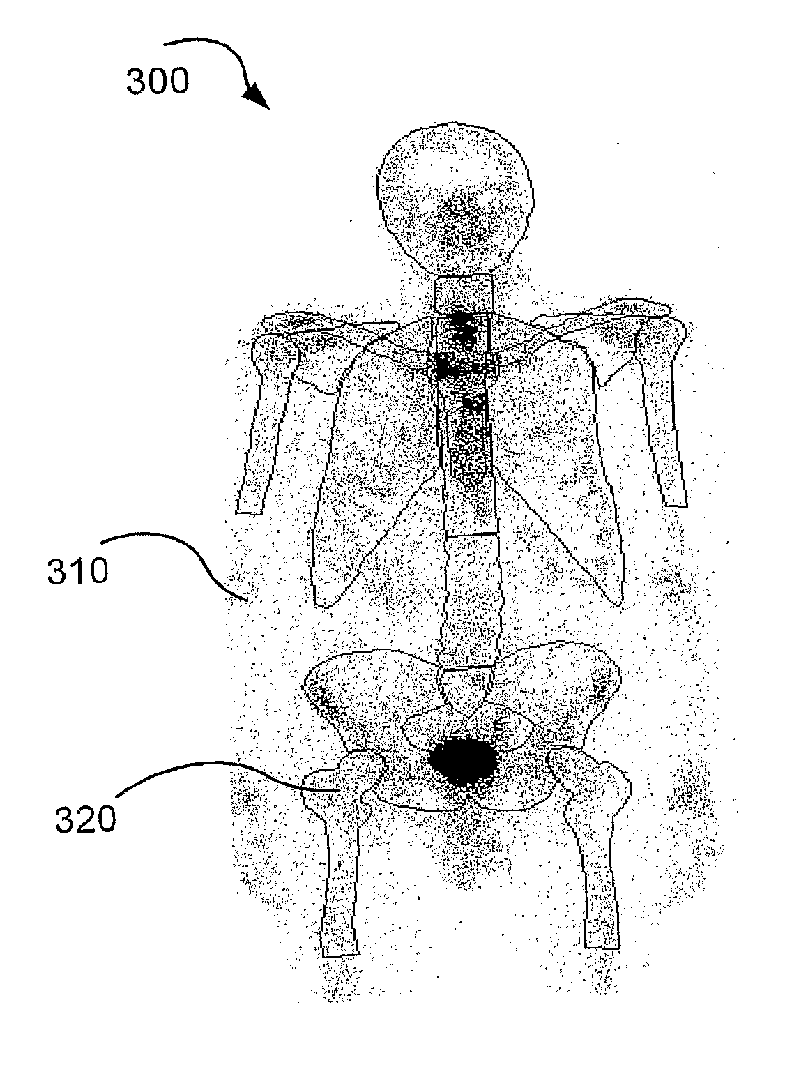
[0039]Embodiments of the present invention relate, in general, to the field of medical imaging and to the field of automated processing and interpretation of medical images. A preferred embodiment relates to a method for automatically or semi-automatically determining contours of a human skeleton and any cancer metastases contained therein and being capable of extracting features to be used by an automatic interpretation system

[0040]An image is a digital representation wherein each pixel represents a radiation intensity, a so called “count”, as known in the art, coming from a radio active substance injected into the human body prior to taking of the image.

[0041]Embodiments of the present invention will be described more fully hereinafter with reference to the accompanying drawings, in which embodiments of the invention are shown. This invention may, however, be embodied in many different forms and should not be construed as limited to the embodiments set forth herein. Rather, these ...


PUM


Abstract

The invention relates to a detection system for automatic detection of bone cancer metastases from a set of isotope bone scan images of a patients skeleton, the system comprising a shape identifier unit, a hotspot detection unit, a hotspot feature extraction unit, a first artificial neural network unit, a patient feature extraction unit, and a second artificial neural network unit.

Description

TECHNICAL FIELD[0001]The present invention relates to the field of medical imaging and to the field of automated processing and interpretation of medical images. In particular, it relates to automated processing and interpretation of two-dimensional bone scan images produced via isotope imaging.BACKGROUND[0002]Interpreting medical images originating from different types of medical scans is a difficult, error prone, and time consuming work which often involves several manual steps. This is especially true when trying to determining contours of a human skeleton and cancer metastases in a medical scan image.[0003]Therefore, there is a great need for a method for determining contours of a human skeleton and any cancer metastases, and being capable of extracting features for an automatic interpretation system.SUMMARY OF THE INVENTION[0004]With the above and following description in mind, then, an aspect of the present invention is to provide a method for determining contours of a human s...

Claims


Application Information

Patent Timeline
Patent Type & Authority Applications(United States)
IPC IPC(8): A61B6/00A61B5/107G06K9/46
CPCA61B6/12G06T2207/10128G06K2209/055G06N3/02G06T7/0012G06T7/0081G06T7/0089G06T2207/20084G06T2207/20124G06T2207/20128G06T2207/20148G06T2207/30004G06T2207/30008G06T2207/30096A61B5/1079A61B6/48G06K9/46A61B5/7264A61B5/7267A61B6/505G06T7/11G06T7/149G06T7/136G16H50/20G06V2201/033
Inventor HAMADEH, IMANNORDBLOM, PIERRESJOSTRAND, KARL
Owner EXINI DIAGNOSTICS
Why Eureka
Try Eureka