Classification of biological tissue by multi-mode data registration, segmentation and characterization

a multi-mode data and biological tissue technology, applied in the field of biological tissue classification by multi-mode data registration, segmentation and characterization, can solve the problems of ineffective targeted therapies and less effective x-ray mammography in tn breast cancer screening than dce-mri, and achieve the effect of reducing the cost of x-ray mammography

Active Publication Date: 2013-08-08
ELUCID BIOIMAGING INC +3
View PDF2 Cites 48 Cited by
  • Summary
  • Abstract
  • Description
  • Claims
  • Application Information

AI Technical Summary

Problems solved by technology

Because of its lack of receptor expression, targeted therapies are ineffective, and chemotherapy is currently the only treatment available.
Studies have shown that x-ray mammography is less effective in TN breast cancer screening than DCE-MRI due to the greater sensitivity of DCE-MRI in detecting TN and basal-like breast cancer phenotypes.

Method used


Image

  • Classification of biological tissue by multi-mode data registration, segmentation and characterization
  • Classification of biological tissue by multi-mode data registration, segmentation and characterization
  • Classification of biological tissue by multi-mode data registration, segmentation and characterization

Examples


Embodiment Construction

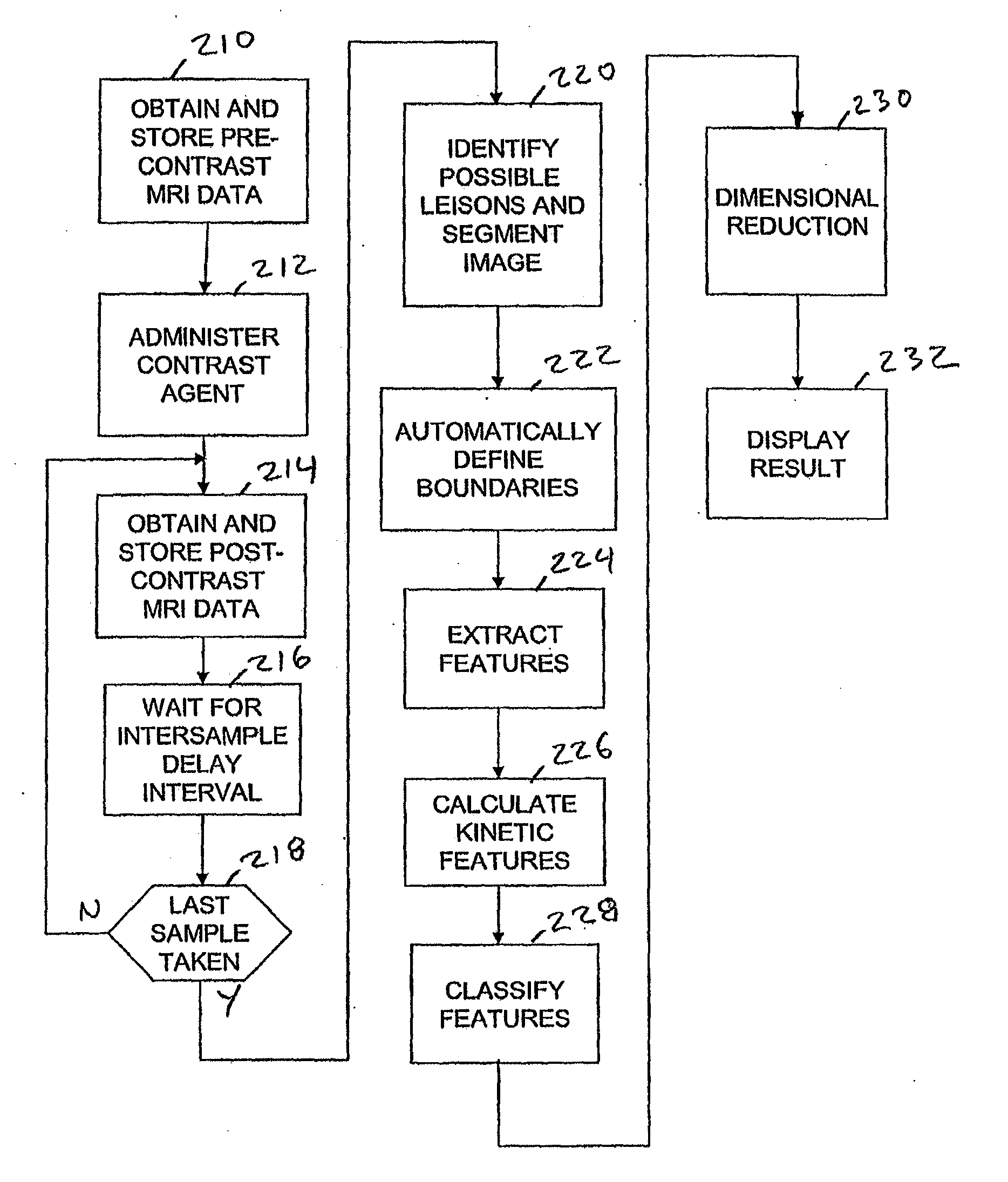
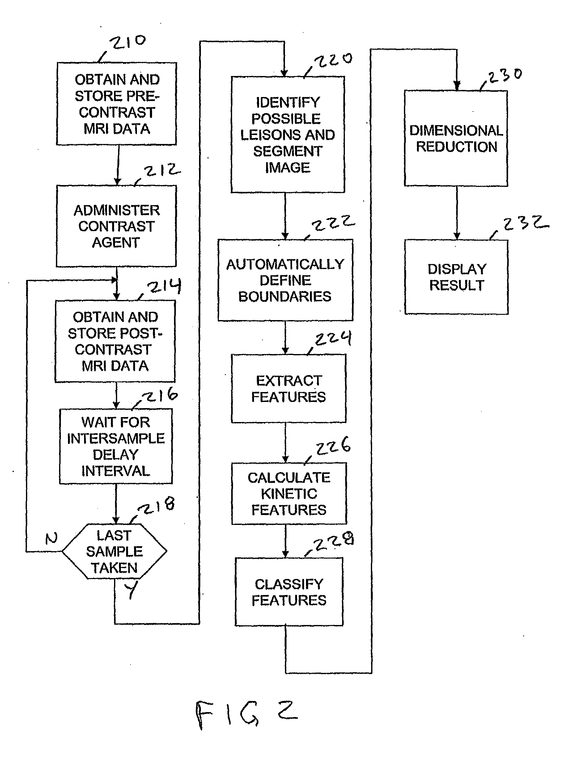
[0016]While the embodiments of the subject invention described below concern the detection of breast tumors, it is contemplated that they may be applied generally to detecting and classifying possibly malignant lesions in other parts of the body based on DCE-MRI data. In addition, the invention may be applied to detect and classify non-malignant regions of interest (ROI) in a body.

[0017]For example, cardiovascular disease is one of the leading causes of death in the United States and world-wide. Atherosclerotic plaques are implicated in many acute cardiovascular events. Furthermore, asymptomatic vulnerable plaques may provide an even greater risk for these acute cardiovascular events. For example, stroke is currently the third largest killer in the United States, with millions dying every year. A more alarming statistic is that about 50% of women and about 64% of men who die from an acute atherosclerotic stroke report no prior symptoms.

[0018]Asymptomatic plaques, also known as vulne...


PUM


Abstract

A method and apparatus for classifying possibly vulnerable plaques from sets of DCE-MRI images includes receiving a set of MRI slice images obtained at respectively different times, where each slice image includes voxels representative of at least one region of interest (ROI). The images are processed to determine the boundaries of the ROIs and the voxels within the identified boundaries in corresponding regions of the images from each time period are processed to extract kinetic texture features. The kinetic texture features are then used in a classification process which classifies the ROIs as vulnerable or stable.

Description

[0001]The present application is a Continuation in Part application of U.S. application Ser. No. 12 / 867,349 filed Oct. 19, 2010, which claims the benefit of PCT International application no. PCT / US2009 / 034505 filed Feb. 19, 2009, which claims the benefit of priority from U.S. provisional application No. 61 / 029,697 filed Feb. 19, 2008 the contents of which are incorporated herein by reference.STATEMENT REGARDING FEDERALLY SPONSORED RESEARCH[0002]The present invention was supported in part by a grant from the National Institutes of Health (NIH) (Grant No. P50 HL083801). The United States Government has certain rights to the invention.BACKGROUND OF THE INVENTION[0003]Magnetic resonance imaging (MRI) of the breast is quickly becoming an important component of breast cancer screening in the United States. The American Cancer Society revised its MRI screening guidelines in 2007 to include women with a 20 to 25 percent increased lifetime risk, including family history of breast and ovarian...

Claims


Application Information

Patent Timeline
Patent Type & Authority Applications(United States)
IPC IPC(8): G06T7/00
CPCG06T7/0012G06T7/0083G06T7/0087G06T2207/30068G06T2207/10096G06T2207/20116G06T7/0089G06T7/12G06T7/143G06T7/149
Inventor BUCKLER, ANDREWMADABHUSHI, ANANTHAMILTON, JAMESAGNER, SHANNONROSEN, MARK
Owner ELUCID BIOIMAGING INC
Why Eureka
Try Eureka