
Augmented reality apparatus

a technology of augmented reality and apparatus, applied in the field of augmented reality, can solve the problems of inability to display images, difficulty in interpolating and registering images to the patient's physical space, and relative cost of integral videography display,

Inactive Publication Date: 2014-01-23
UNIV HEALTH NETWORK
View PDF5 Cites 424 Cited by
  • Summary
  • Abstract
  • Description
  • Claims
  • Application Information

AI Technical Summary

Benefits of technology

The present patent relates to an apparatus and method for providing assistance to intervention procedures such as surgery and needle biopsy. The apparatus uses augmented reality to superimpose patient-specific medical image information on the intervention site. The apparatus includes a trackable projector that projects an image based on the position and orientation of the intervention site and the tracked position of the projector. The apparatus also has a data processor and a tracking device that provide data to the processor to determine the spatial relationship between the projector and the intervention site. Using this information, the processor generates data representative of the medical image to be projected by the projector and instructs it to project the image. The technical effect of the patent is that it provides a more precise and accurate guidance for intervention procedures and improves the accuracy and precision of surgery and needle biopsy.

Problems solved by technology

With such an arrangement, the clinician's sight must be directed away from the operation field in order to view the monitor, which may result in difficulties for the clinician in interpolating and registering the image to the patient's physical space.
The need for such mental interpolation and registration may lead to complications, for example if the clinician is not familiar with correlation of guidance image to the operation site or if the clinician lacks sophisticated hand-eye coordination training for the specific procedure.
However, such integral videography display can be relative expensive and not commonly available.
However, such systems are non-mobile, may require the imaging device to be fixed in-place, and may be unable to display images that do not align with the imaging plane of the scanner.
A common drawback of HMD can include an indirect vision of the operating field, the need for extra equipment weight on the user's head thereby preventing long-time usage, and restricted user motion due to wiring associated with the head-mounted display.

Method used


Image

  • Augmented reality apparatus
  • Augmented reality apparatus
  • Augmented reality apparatus

Examples


Embodiment Construction

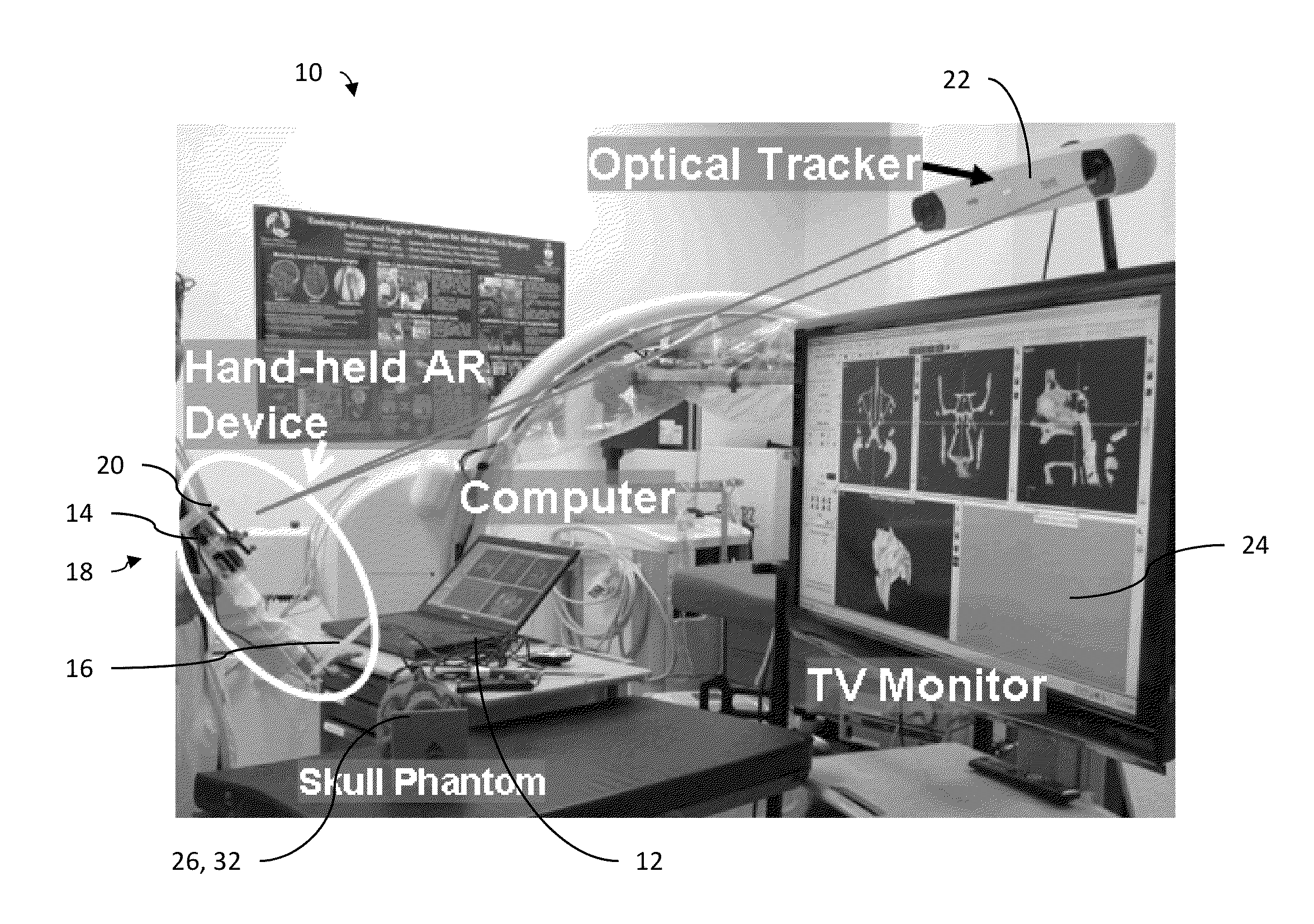
[0028]The design and implementation of mobile handheld augmented reality apparatus and systems for image guidance suitable for use in intervention procedures are disclosed herein. Aspects of various embodiments are described below through reference to the drawings. An exemplary embodiment of a handheld augmented reality (AR) apparatus may be relatively compact and mobile, and may allow for medical imaging and / or planning data to be spatially superimposed on a visual target of interest, such as an intervention (e.g., surgical) site. In some examples, the AR apparatus may include a micro-projector, translucent display (also referred herein as a head-up display), computer interface, camera, instrument tracking system, head tracking system and dedicated software for real-time tracking, navigation and visualization (also referred herein as “X-Eyes”).

[0029]In some aspects, the disclose apparatus may provide one or more functionalities to augment viewing of an intervention site, such as by...


PUM


Abstract

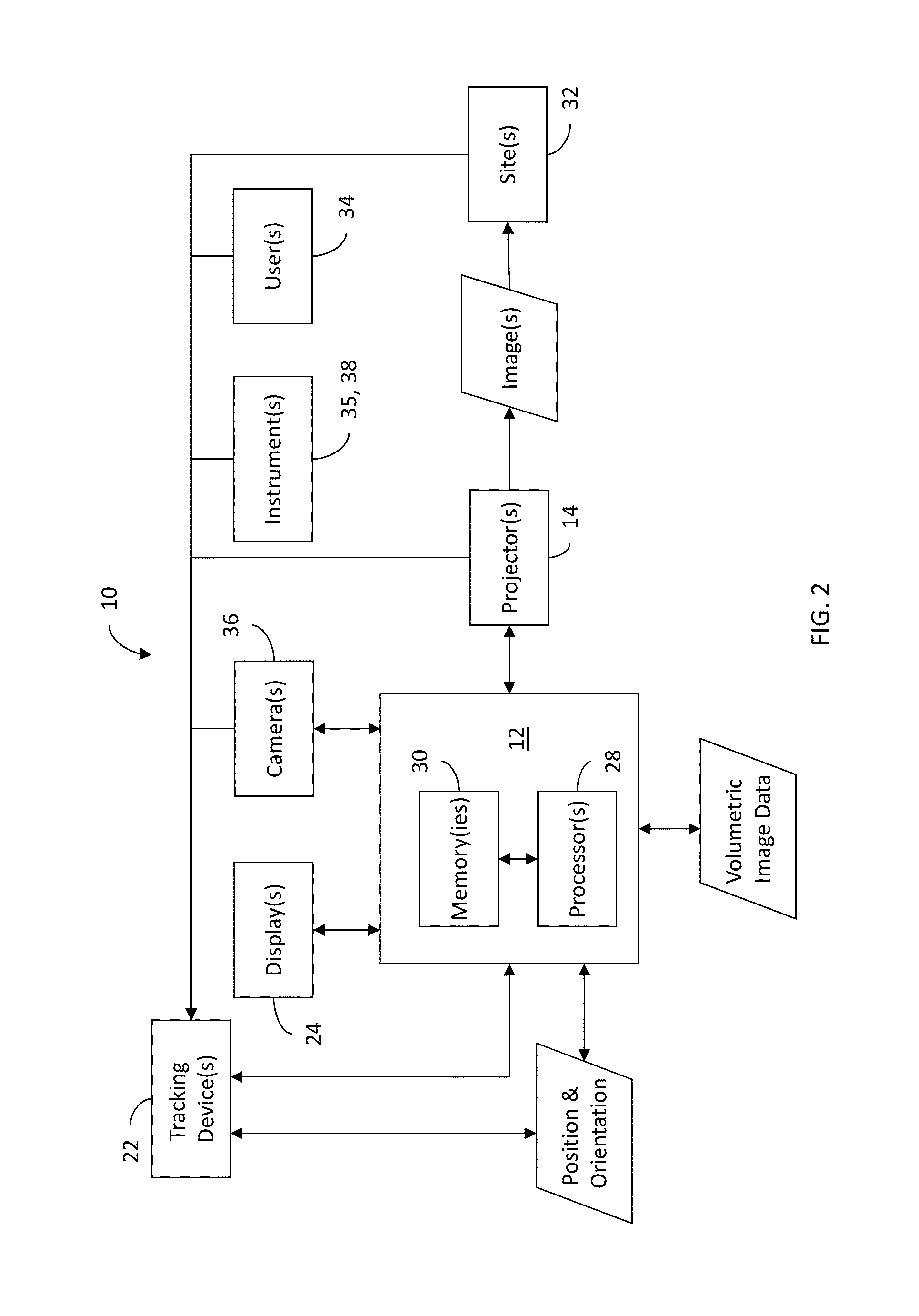
Augmented reality apparatus (10) for use during intervention procedures on a trackable intervention site (32) are disclosed herein. The apparatus (10) comprises a data processor (28), a trackable projector (14) and a medium (30) including machine-readable instructions executable by the processor (28). The projector (14) is configured to project an image overlaying the intervention site (32) based on instructions from the data processor (28). The machine readable instructions are configured to cause the processor (28) to determine a spatial relationship between the projector (14) and the intervention site (32) based on a tracked position and orientation of the projector (14) and on a tracked position and orientation of the intervention site (32). The machine readable instructions are also configured to cause the processor to generate data representative of the image projected by the projector based on the determined spatial relationship between the projector and the intervention site.

Description

CROSS REFERENCE TO RELATED APPLICATION AND CLAIM OF PRIORITY[0001]The present application claims priority to U.S. provisional patent application No. 61 / 673,973 filed on Jul. 20, 2012, the entire contents of which are hereby incorporated by reference.TECHNICAL FIELD[0002]The disclosure relates generally to augmented reality and more particularly to augmented reality apparatus and methods for use in medical and related applicationsBACKGROUND OF THE ART[0003]Conventional image-guided systems have been used to enable clinicians to navigate medical instruments advancing in patient space during intervention procedures. Such systems typically involve the use of a monitor displaying medical images to guide the clinician to a target. Typically, the monitor is arranged in a stationary position, such as in-place on a desk or wall-mounted close to the patient's bed. With such an arrangement, the clinician's sight must be directed away from the operation field in order to view the monitor, which...

Claims


Application Information

Patent Timeline
IPC IPC(8): G09G5/377
CPCG09G5/377A61B2034/2055A61B2090/363A61B2090/366A61B2090/372G02B27/017G02B2027/0138G02B2027/014G02B2027/0187G06F3/011G06F3/147G09G3/002G09G2340/12G09G2354/00G09G2380/08G16H30/20H04N9/3185
Inventor CHAN, HARLEY HAU-LAMDALY, MICHAEL JOHNIRISH, JONATHAN CRAWFORD
Owner UNIV HEALTH NETWORK
Learn More