
X-Ray Identification for Active Implantable Medical Device

a medical device and active technology, applied in the field can solve the problems of more difficult to find acceptable locations and more difficult to radiograph the identification of implantable medical devices

Active Publication Date: 2014-02-27
PACESETTER INC
View PDF3 Cites 129 Cited by
  • Summary
  • Abstract
  • Description
  • Claims
  • Application Information

AI Technical Summary

Benefits of technology

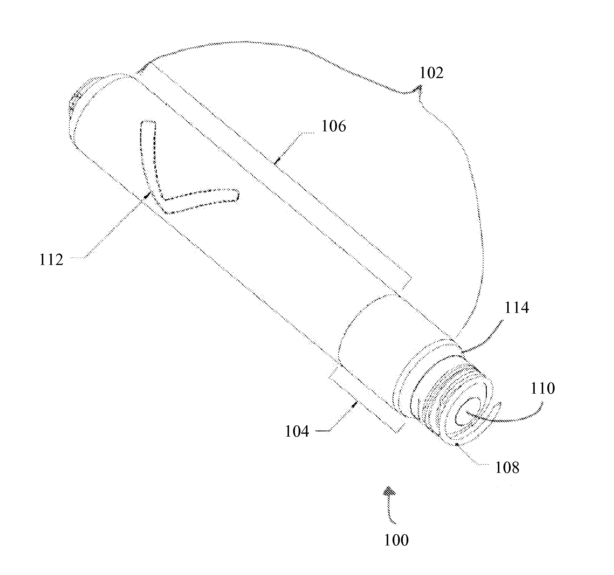
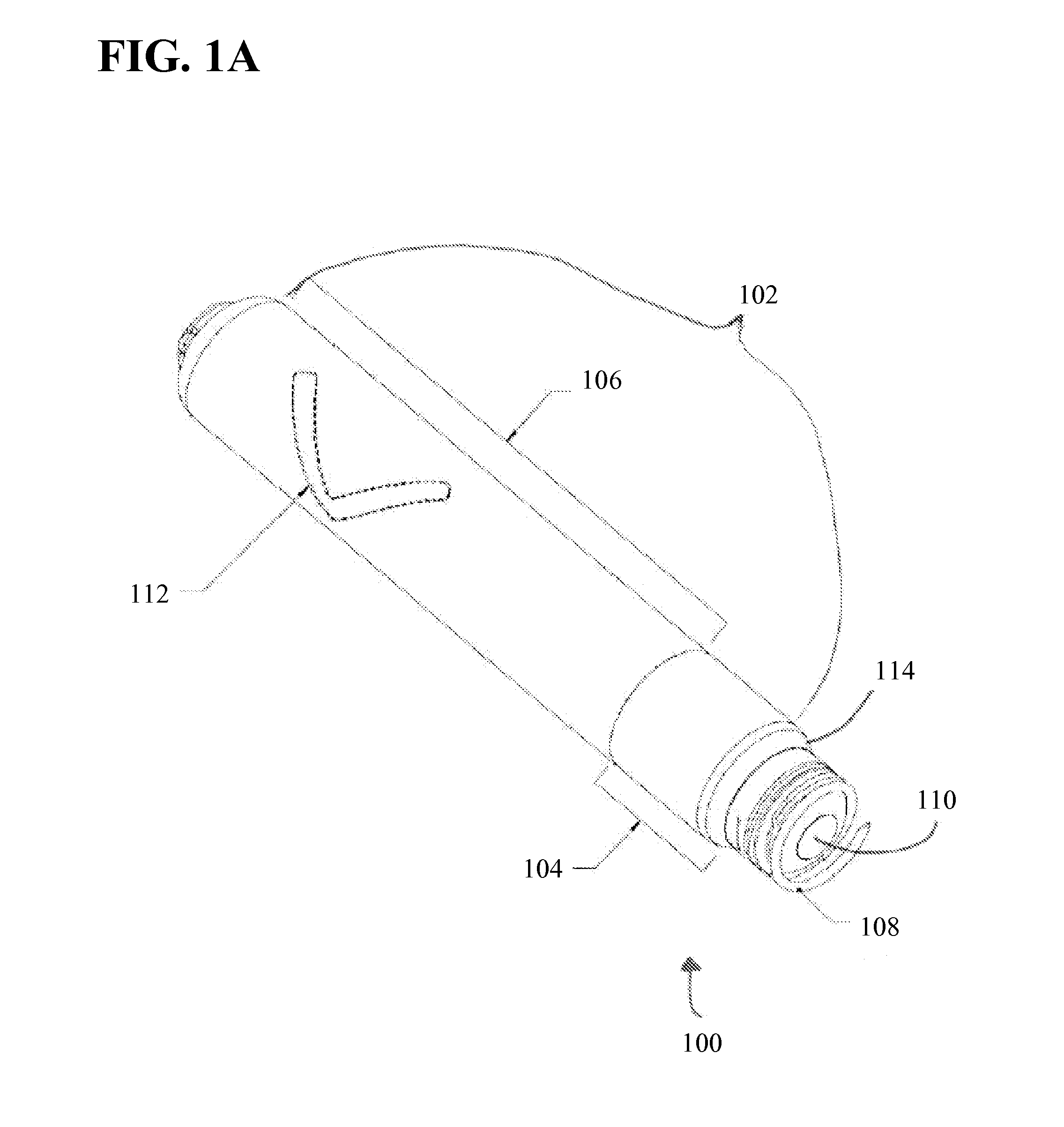
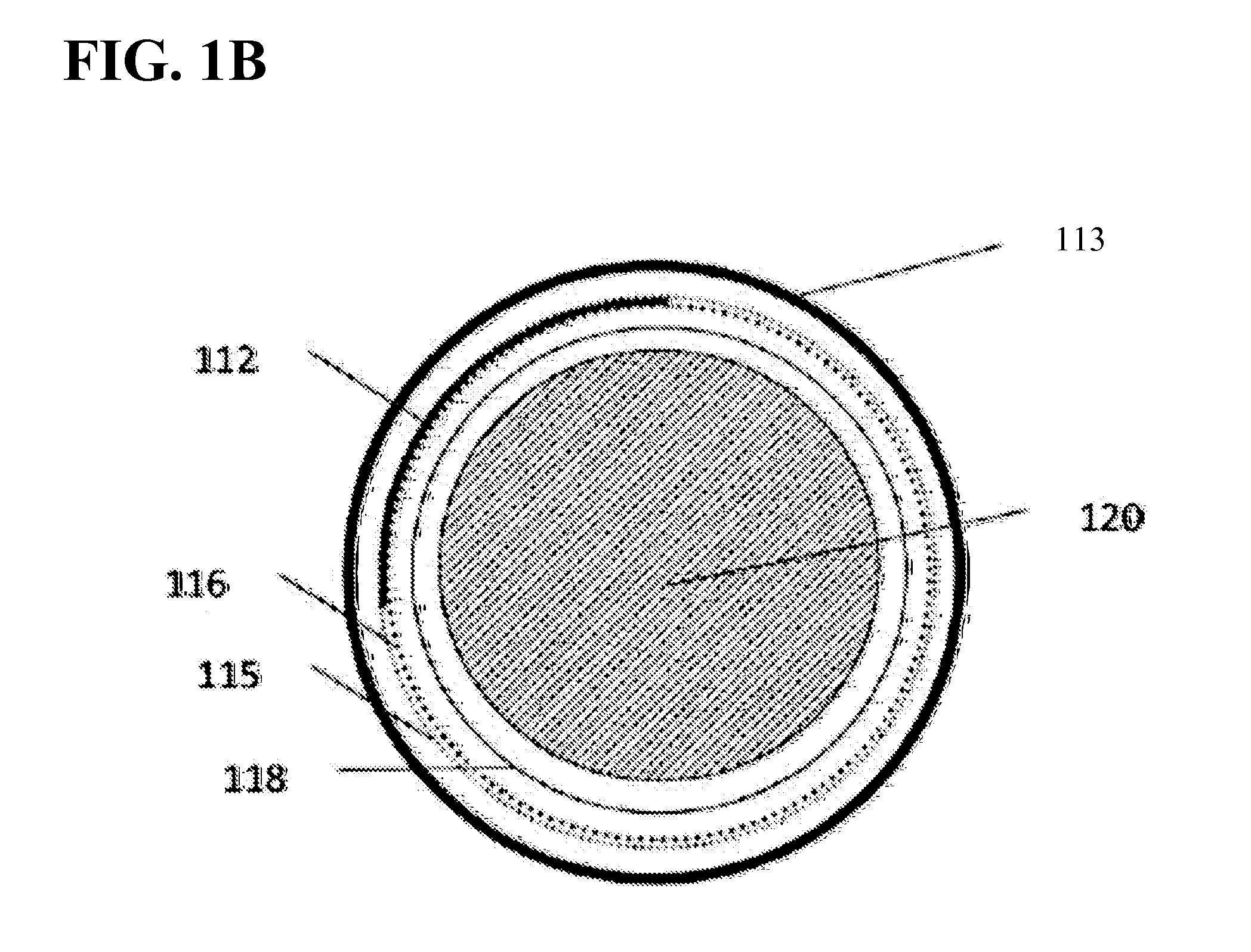
This patent describes an implantable medical device, such as a leadless pacemaker, that has a cell and a radio-opaque marker integrated into the cell. The radio-opaque marker can be located inside or outside of the cell and can be made of various materials like tungsten, tantalum, platinum, or gold. The device can be shaped to indicate its rotation when scanned by X-ray radiation. The device also has a smooth exterior surface, and a thermal separator can be used between the energy source and the cell. The technical effects include improved visualization of the implant and more accurate placement of the pacing electrodes.

Problems solved by technology

Radiographic identification of implantable medical devices has become more challenging because of the reduction in volume and size of implantable medical devices.
The reduction in size of implantable medical devices makes it more challenging to find acceptable locations for placing radiographic tags that can be identified and visualized using standard medical radiographic equipment.

Method used


Image

  • X-Ray Identification for Active Implantable Medical Device
  • X-Ray Identification for Active Implantable Medical Device
  • X-Ray Identification for Active Implantable Medical Device

Examples


Embodiment Construction

[0035]Technological advances have allowed for a reduction in the size of active implantable medical devices. Decreasing the size of the implantable medical devices has made it more challenging to find acceptable locations for placing radiographic markers or tags on the active medical device. Radiographic visualization of the implantable medical device can be more difficult because of limits with conventional X-ray machines and the orientation of the body of the patient with respect to conventional X-ray machines.

[0036]Active implantable medical devices typically have an electronics compartment and a cell. The radiographic or radio-opaque marker or tag is usually placed inside the electronics compartment. However, the electronics compartment has radio-opaque components such as tantalum capacitors. The radio-opaque tag or marker can be shadowed, obscured, or confused with the neighboring radio-opaque materials using conventional fluoroscopy methods. The components of the electronics c...


PUM


Abstract

An active implantable medical device is disclosed herein having a radio-opaque marker. The radio-opaque marker can be formed within an exterior wall of the device or within recesses on the outside of the exterior wall. The implantable medical device can be a leadless pacemaker. The shape of the radio-opaque marker can be designed to facilitate visualization and identification of the location, orientation, and rotation of the implanted medical device by conventional fluoroscopy. Methods of use are also disclosed.

Description

CROSS-REFERENCE TO RELATED APPLICATIONS[0001]This application claims the benefit of U.S. Provisional Application No. 61 / 691,721, filed on Aug. 21, 2012, titled “X-Ray Identification for Active Implantable Medical Device”, the contents of which are incorporated by reference herein.INCORPORATION BY REFERENCE[0002]All publications and patent applications mentioned in this specification are herein incorporated by reference to the same extent as if each individual publication or patent application was specifically and individually indicated to be incorporated by reference.FIELD[0003]This disclosure generally relates to implantable medical devices and more particularly to implantable leadless cardiac pacemakers.BACKGROUND[0004]Radiographic identification has been useful in helping clinicians identify in-vivo active implantable medical devices, such as implantable cardiac pacemakers. Typically, identification occurs via X-ray illumination to observe a radio-opaque tag or marker located ins...

Claims


Application Information

Patent Timeline
Patent Type & Authority Applications(United States)
IPC IPC(8): A61N1/05
CPCA61N1/0587A61N1/3756A61N1/0573A61N1/37205A61B2090/3966
Inventor OSTROFF, ALANPASPA, PAULJACOBSON, PETER M.KELLER, WADE A.HUBBARD, CHRISTOPHER ALAN
Owner PACESETTER INC
Learn More