Region extraction apparatus, method and program

a technology of extraction apparatus and region, applied in image analysis, image enhancement, instruments, etc., can solve the problems of long processing time and higher risk of error detection, and achieve the effect of limiting the range of extraction processing and high-accuracy extraction processing

Active Publication Date: 2014-03-20
FUJIFILM CORP
View PDF12 Cites 17 Cited by
  • Summary
  • Abstract
  • Description
  • Claims
  • Application Information

AI Technical Summary

Benefits of technology

[0037]According to the region extraction apparatus, method and program of the present invention, a bronchial structure representing a structure of a bronchus or bronchi is extracted from a three-dimensional medical image of a chest. The extracted bronchial structure is divided, based on the divergence of the bronchial structure, into a plurality of divided bronchial structures. A plurality of divided lung regions are obtained based on the plurality of divided bronchial structures. A distance image based on a distance between each voxel in an entire region excluding at least one of the plurality of divided lung regions and each of th...

Problems solved by technology

Therefore, there are problems that the time period of extraction ...

Method used


Image

  • Region extraction apparatus, method and program
  • Region extraction apparatus, method and program
  • Region extraction apparatus, method and program

Examples


first embodiment

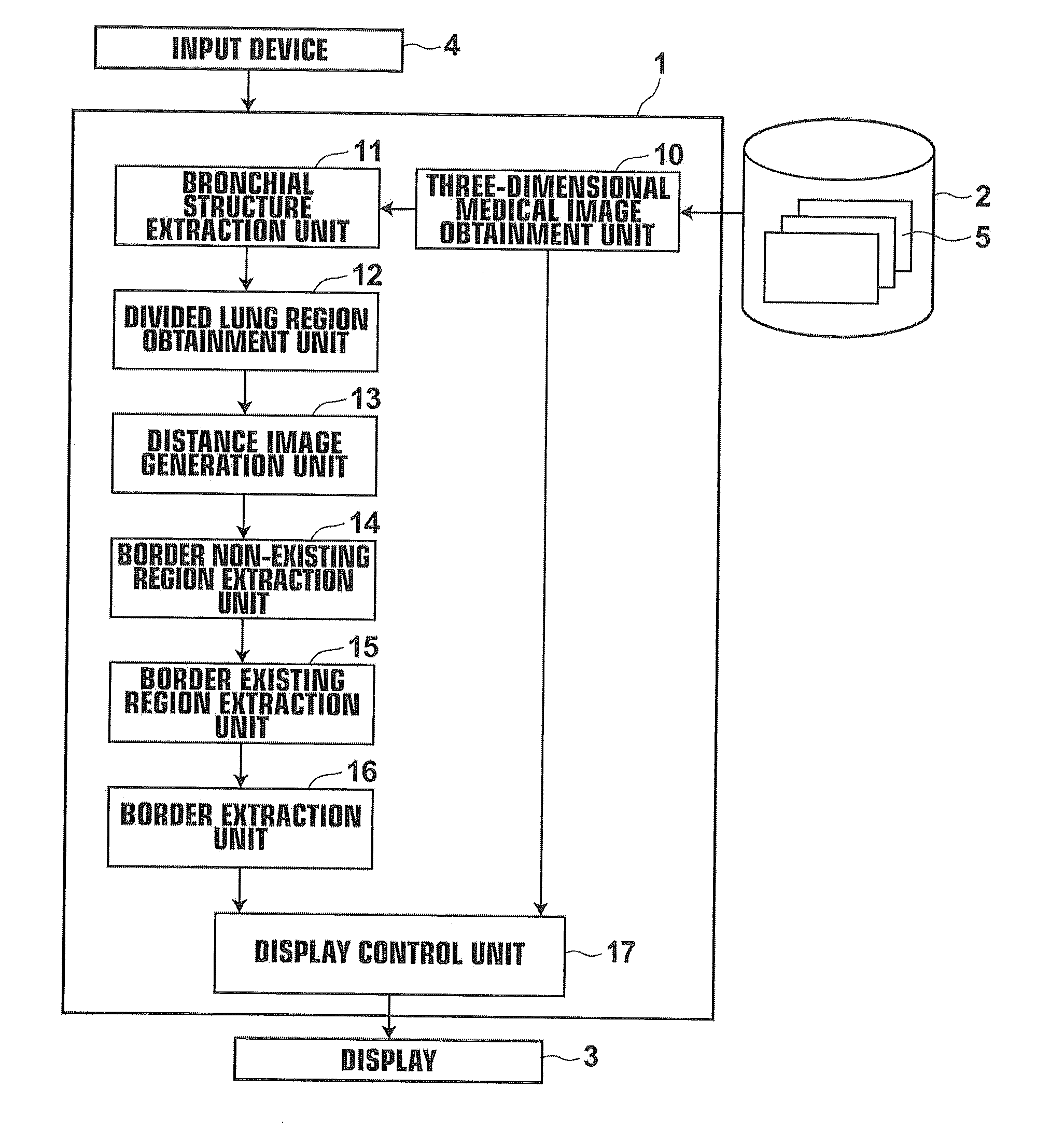
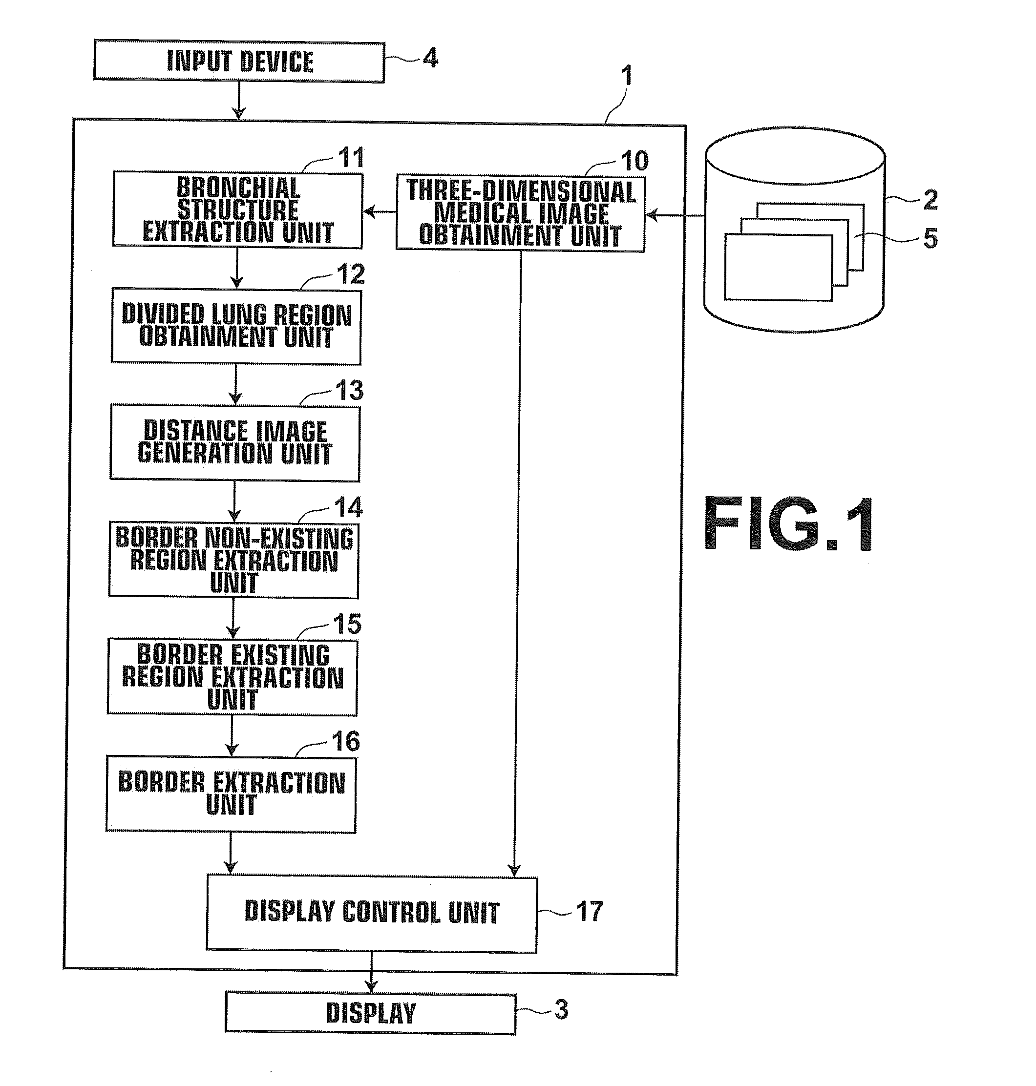
[0054]Hereinafter, a medical image diagnosis support system using a region extraction apparatus, method and program of the present invention will be described in detail with reference to drawings. FIG. 1 is a schematic block diagram illustrating the configuration of a medical image diagnosis support system according to an embodiment of the present invention. The medical image diagnosis support system according to the embodiment of the present invention extracts a lung region from a three-dimensional medical image of the chest of a subject (patient) to be examined. The medical image diagnosis support system extracts interlobar fissures, as illustrated in FIG. 2, from the lung region at high accuracy and at high speed, and displays the extracted interlobar fissures.

[0055]The medical image diagnosis support system according to an embodiment of the present invention includes a medical image display apparatus 1, a three-dimensional medical image storage server 2, a display 3, and an inpu...

second embodiment

[0108]Specifically, as illustrated in FIG. 11, first, the distance image generation unit 13 leaves only right upper lobe region RA of the three lung lobe regions in entire region R of the right lung. Further, the distance image generation unit 13 calculates distance DA between each voxel P in the entire region R excluding the right upper lobe region RA and the right upper lobe region RA. Further, the distance image generation unit 13 generates distance image A′, as one of third distance images, by assigning the value of the distance DA to each voxel P.

[0109]Then, as illustrated in FIG. 12, the distance image generation unit 13 leaves only right middle lobe region RB of the three lung lobe regions in entire region R of the right lung. The distance image generation unit 13 calculates distance DB between each voxel P in the entire region R excluding the right middle lobe region RB and the right middle lobe region RB. Further, the distance image generation unit 13 generates distance im...


PUM


Abstract

A three-dimensional medical image obtainment unit that obtains a three-dimensional medical image of a chest, a bronchial structure extraction unit that extracts a bronchial structure from the three-dimensional medical image, a divided lung region obtainment unit that divides, based on the divergence of the bronchial structure, the bronchial structure into plural bronchial structures, and obtains plural divided lung regions based on the plural divided bronchial structures, a distance image generation unit that generates, based on the plural divided lung regions, a distance image based on a distance between each voxel in an entire region excluding at least one of the plural divided lung regions and each of the plural divided lung regions, and a border non-existing region extraction unit that extracts, based on the distance image generated by the distance image generation unit, a border non-existing region, which does not include any borders of the divided lung regions, are provided.

Description

BACKGROUND OF THE INVENTION[0001]1. Field of the Invention[0002]The present invention relates to a region extraction apparatus, method and program for extracting a region, such as a lung lobe, from a three-dimensional medical image of a chest. In particular, the present invention relates to a region extraction apparatus, method and program for extracting a border of the region, such as a lung lobe.[0003]2. Description of the Related Art[0004]In a surgery of a lung cancer or the like, it is important to recognize interlobar fissures, which are present between lung lobe regions, and borders of lung segments to conserve pulmonary functions after the surgery and to maintain cardiopulmonary functions.[0005]Conventionally, such information about interlobar fissures and borders of lung segments was displayed, for example, by directly inputting the information by a manual operation, or by obtaining interlobar fissures by manual input of each lung lobe region.[0006]Meanwhile, as image recogn...

Claims


Application Information

Patent Timeline
IPC IPC(8): G06T7/00
CPCG06T7/0083G06T2207/10081G06T2207/10088G06T2207/20021G06T2207/30061G06T2207/30064G06T7/12
Inventor INOUE, TSUTOMU
Owner FUJIFILM CORP
Why Eureka
Try Eureka