Method and system of measuring anatomical features in subcutaneous images to assess risk of injury

a technology of anatomical features and subcutaneous images, applied in the field of methods and systems of measuring anatomical features in subcutaneous images to assess risk of injury, can solve the problems of inability to detect injury to occ ligaments, progress quickly and catastrophically, and spinal cord injury and death, and achieve the effect of effective indicator of ligament injury

Inactive Publication Date: 2014-05-15
SCOTT & WHITE HEALTHCARE +1
View PDF0 Cites 17 Cited by
  • Summary
  • Abstract
  • Description
  • Claims
  • Application Information

AI Technical Summary

Benefits of technology

[0015]The present disclosure provides a method and system using software and associated equipment to automatically identify two or more preselected anatomical features by examining pixels within images such as computerized tomography (CT) scanned images, determining appropriate measurements between such identified features based on dimensions and scaling available within the image file, comparing these measurements to normative data, and providing output of the results to a user, such as medical personnel. In at least one embodiment, the system can compute the basion-dens interval (BDI), which has been shown to be an effective indicator of ligament injury within the occipito-cervical complex (“OCC”) that may not present with other symptoms, is not revealed with standard CT imaging, and is of mortal danger to the patient if untreated. The system can automate interpreting CT scanned images and alert medical personnel about the risk of OCC ligament damage.

Problems solved by technology

High-energy deceleration force often associated with motor vehicle collisions can compromise the stability provided by the strong OCC ligaments and result in spinal cord injury and death.
Some of these patients present neurologically intact, but if the OCC damage is not quickly and properly diagnosed and treated it can progress quickly and catastrophically.
Injury to the OCC ligaments can be difficult to detect.
However, accurately identifying the skeletal landmarks and properly interpreting the scans requires unusual care, training, and expertise.
If this distance exceeds a certain threshold, then the potential risk of undetected OCC injury is substantially elevated.
Such a manual determination, however, is tedious, time-consuming, and susceptible to human error, despite the highest level of medical training, skill, and experience.

Method used


Image

  • Method and system of measuring anatomical features in subcutaneous images to assess risk of injury
  • Method and system of measuring anatomical features in subcutaneous images to assess risk of injury
  • Method and system of measuring anatomical features in subcutaneous images to assess risk of injury

Examples


example

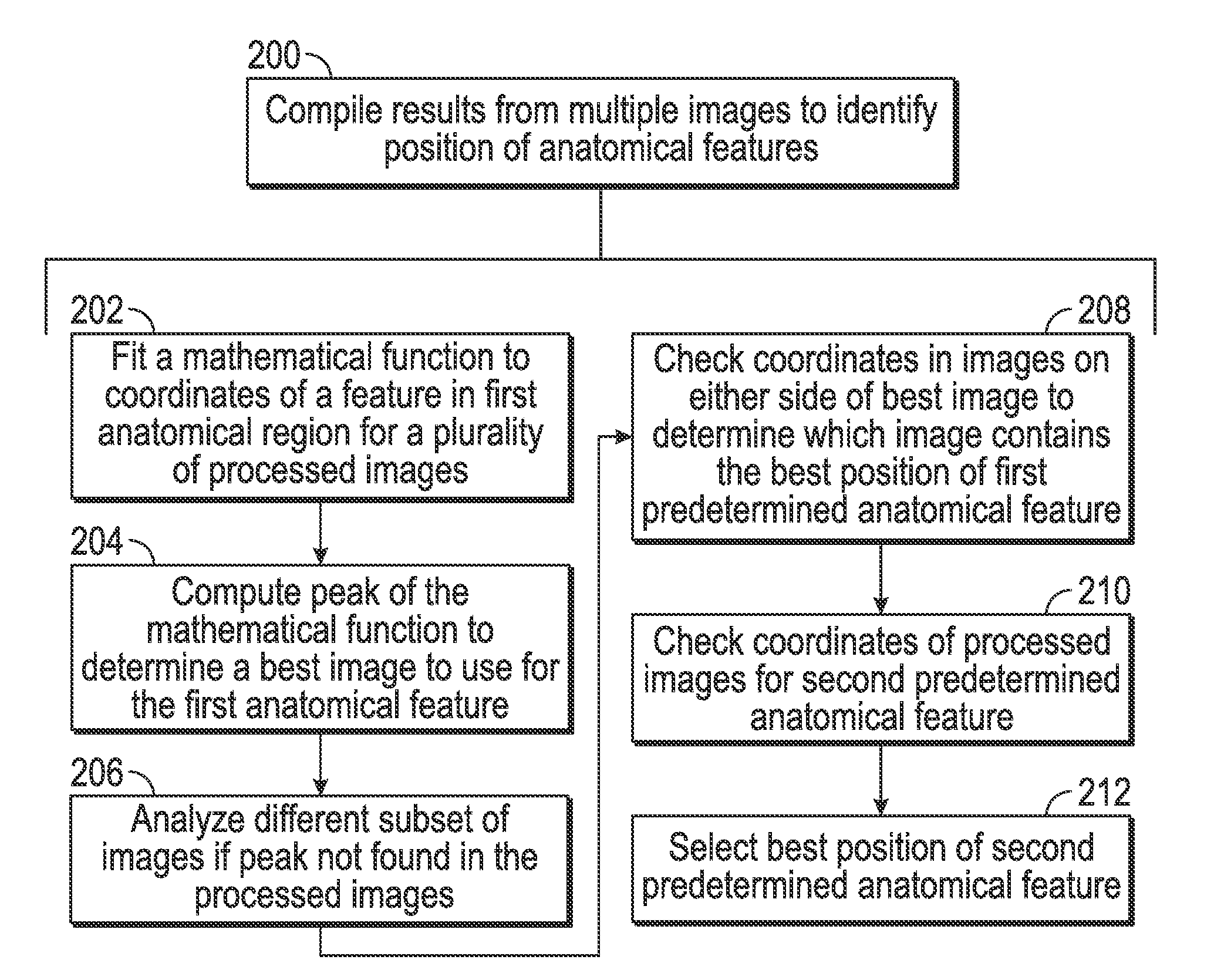
[0100]While the above explanation has been generalized for anatomical features with some discussion of the BDI measurement, the following description provides more details and specific information for the BDI measurement and comparison to normative data for an assessment of the risk for OCC injury. The principles, logic, and considerations, such as starting points in images and stepwise process for directional movements in analyzing pixels, which are provided in this example, can be applied to other sequences and other anatomical features. Thus, the details in this example are only exemplary and not limited to these particular anatomical features, and are explicitly intended to serve as guidelines for other sequences and anatomical features using the teachings herein.

[0101]Within at least this example, the disclosed system and method, including the software, for determining the BDI accepts as input a series of sagittal-plane CT image files focused on the cervical spine area. It is a...


PUM


Abstract

The present disclosure provides a method and system using software and associated equipment to automatically identify two or more preselected anatomical features by examining pixels within images such as computerized tomography (CT) scanned images, determining appropriate measurements between such identified features based on dimensions and scaling available within the image file, comparing these measurements to normative data, and providing output of the results to a user, such as medical personnel. In at least one embodiment, system can measure the basion-dens interval (BDI), which has been shown to be an effective indicator of ligament injury within the occipito-cervical complex (“OCC”) that may not present with other symptoms, is not revealed with standard CT imaging, and is of mortal danger to the patient if untreated. The system can automate interpreting CT scanned images and alert medical personnel about the risk of OCC ligament damage.

Description

CROSS REFERENCE TO RELATED APPLICATIONS[0001]This application claims the benefit of U.S. Provisional Appl. No. 61 / 724,711 filed on Nov. 9, 2012 and is incorporated herein by reference.STATEMENT REGARDING FEDERALLY SPONSORED RESEARCH OR DEVELOPMENT[0002]Not applicable.REFERENCE TO APPENDIX[0003]Not applicable.BACKGROUND OF THE INVENTION[0004]1. Field of the Invention[0005]The invention disclosed and taught herein relates generally to a method and system of locating and / or tracing along pre-selected anatomical features that includes processing digital images of the general region of the structures to locate the specific features, mathematically determining a distance between such features, comparing the determined distance to normative data, and reporting the results. More specifically, the invention can be used to determine a distance between anatomical features from analyzed subcutaneous images such as computerized tomography (CT) scans, comparing the distance to normative data for ...

Claims


Application Information

Patent Timeline
Patent Type & Authority Applications(United States)
IPC IPC(8): A61B5/00G06T7/00
CPCG06T7/0014A61B5/4504G06T7/0012G06T7/60G06T2207/10081G06T2207/30012A61B6/032A61B6/505A61B6/5217
Inventor GARNER, BRIAN A.CHAPUT, CHRISTOPHERHOFFMAN, JACOB
Owner SCOTT & WHITE HEALTHCARE
Why Eureka
Try Eureka