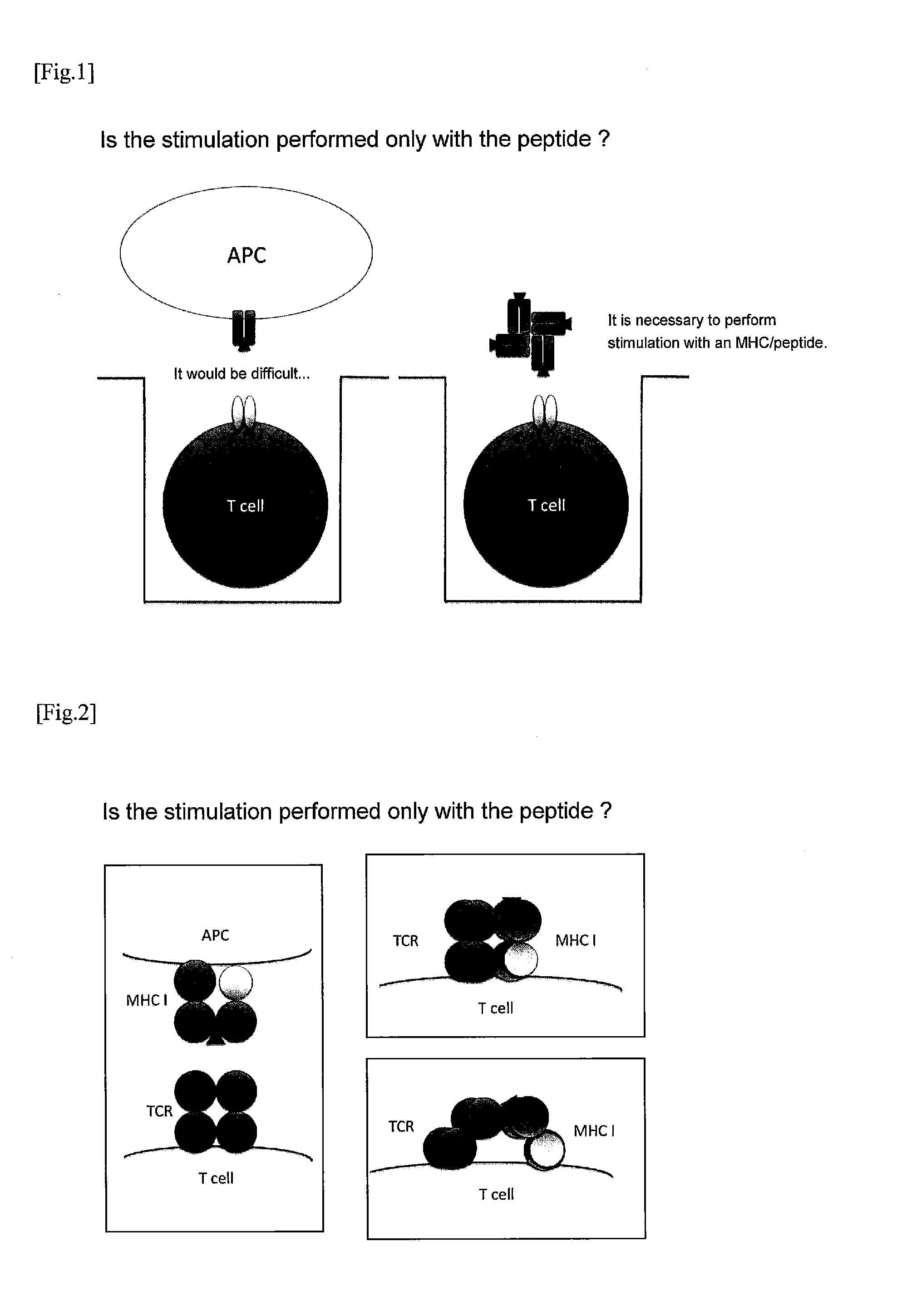
Method for Stimulating T Cell and Use Thereof
a technology of t cell and cell surface, which is applied in the field of method for stimulating and activating a cell, can solve the problems of limited types of currently marketed (mhc/p)/sub>4, and achieve the effect of efficient and convenient identification
- Summary
- Abstract
- Description
- Claims
- Application Information
AI Technical Summary
Benefits of technology
Problems solved by technology
Method used
Image

Examples
example 1
Detection of Antigen-Specific Mouse T Cell
Preparation of T Cells
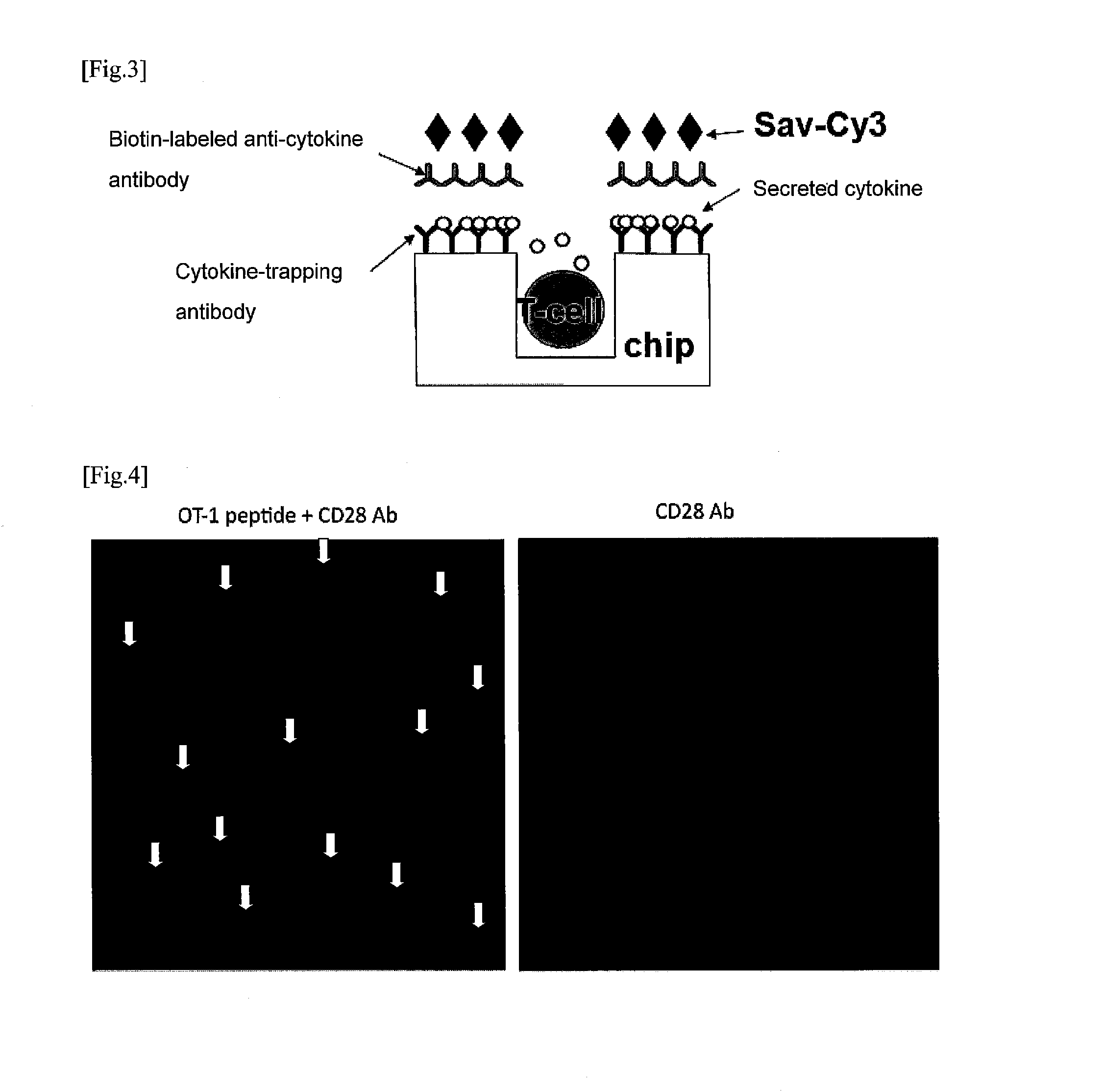
[0102]Lymphocytes of an OT-1 TCR transgenic mouse into which the gene of TCR that recognizes an ovalbumin (OVA)-derived peptide, the OT-1 peptide, was introduced were prepared from the spleen and lymph nodes of the mouse. The lymphocytes were suspended in 10% FCS / RPMI1640 medium at a density of 2.5×10⁶ cells / mL, and used for the following experiments.
Preparation of Chip
[0103]Anti-IL-2 antibodies (5 μg / mL) were put on a microwell array chip (10 μm in diameter, 126,000 wells), and incubated overnight at room temperature to be bound to the chip surface. On the next day, the chip was washed with phosphate buffered saline (PBS), then PBS containing 0.01% Lipidure BL103 (NOF Corporation) was added to the chip surface, and the chip was placed under reduced pressure so that the air in the microwells was evacuated, and Lipidure was contacted with the chip surface and internal surfaces of the wells to perform blocking (room tempe...
example 2
Detection of Antigen-Specific Mouse T Cell
Preparation of T Cells
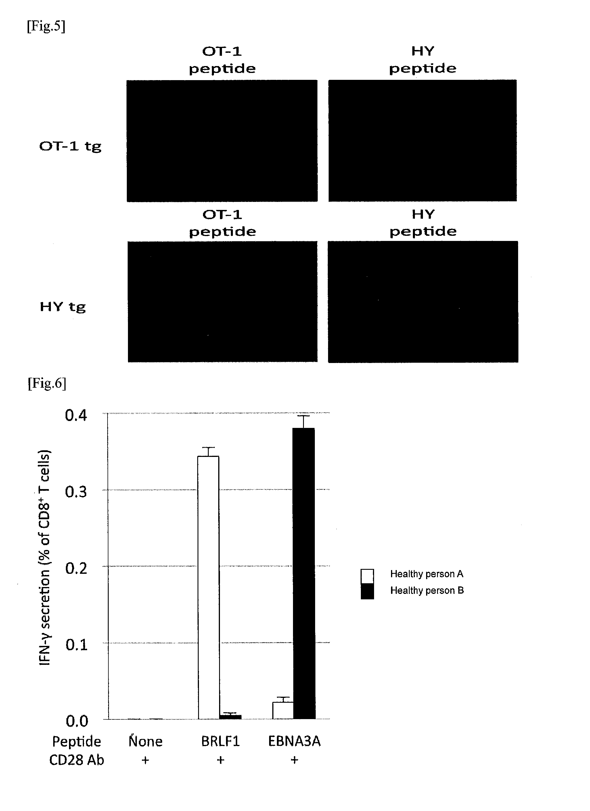
[0107]Lymphocytes of an H-Y TCR transgenic mouse into which the gene of TCR that recognizes an H-Y antigen-derived peptide, which is specifically expressed in male, was introduced, and lymphocytes of an OT-1 TCR transgenic mouse into which the gene of TCR that recognizes an ovalbumin (OVA)-derived peptide, the OT-1 peptide, was introduced were prepared from the spleens and lymph nodes of the mice, respectively. The lymphocytes were suspended in 10% FCS / RPMI1640 medium at a density of 2.5×10⁶ cells / mL, and used for the following experiments.
Preparation of Chip
[0108]The anti-IL-2 antibodies (5 μg / mL) were put on a microwell array chip (10 μm in diameter, 126,000 wells), and incubated overnight at room temperature to be bound to the chip surface. On the next day, the chip was washed with PBS, then PBS containing 0.01% Lipidure BL103 (NOF Corporation) was added to the chip surface, and the chip was placed under reduced pres...
example 3
Detection of Antigen-Specific Human T Cell
Preparation of T Cells
[0113]Most of Japanese are inapparently infected with the Epstein-Barr virus (EB virus). So far, several peptides such as BRLF1 and EBNA3A has been identified as EB virus-derived peptides that bind to the FILA-A24 molecule and serve as a T cell epitope. Peripheral blood was collected from two of healthy persons A and B in a volume of 20 mL each, equal volume of PBS was added to the blood to dilute it 2-fold, and then the diluted blood was layered on Lymphosepar I (Immuno-Biological Laboratories), and centrifuged at 2000 rpm for 30 minutes to separate a lymphocyte fraction. The lymphocytes were suspended in 10% FCS / RPMI1640 medium at a density of 2.5×10⁶ cells / mL, and used for the following experiments.
Preparation of Chip
[0114]Anti-interferon-γ antibodies (5 μg / mL) were put on a microwell array chip (10 μm in diameter, 126,000 wells), and incubated overnight at room temperature to be bound to the chip surface. On the nex...
PUM
| Property | Measurement | Unit |
|---|---|---|
| diameter | aaaaa | aaaaa |
| diameter | aaaaa | aaaaa |
| depth | aaaaa | aaaaa |
Abstract
Description
Claims
Application Information
 - R&D Engineer
- R&D Manager
- IP Professional
- Industry Leading Data Capabilities
- Powerful AI technology
- Patent DNA Extraction
Browse by: Latest US Patents, China's latest patents, Technical Efficacy Thesaurus, Application Domain, Technology Topic.
© 2024 PatSnap. All rights reserved.Legal|Privacy policy|Modern Slavery Act Transparency Statement|Sitemap