Soft tissue lesion excision guide

a soft tissue lesion and guide wire technology, applied in the field of soft tissue lesion excision guide devices, can solve the problems of inadvertent cutting of the lesion, incomplete removal, and missed lesion altogether, and the guide wires currently available are easily dislodged from the lesion

Inactive Publication Date: 2015-02-26
EXCISURE LLC
View PDF5 Cites 7 Cited by
  • Summary
  • Abstract
  • Description
  • Claims
  • Application Information

AI Technical Summary

Benefits of technology

[0012]In a first stored configuration, the encompassing element is inserted into the inner diameter of the insertion element with the curvilinear portions of the shape memory wires having a generally straight configuration. The deployment element is inserted into the proximal end of the inner diameter of the insertion element such that the extension wire resides inside the inner diameter of the deployment element and the distal tip of the deployment element abuts the proximal ends of the three or more shape memory wires. In this configuration, the apparatus may be placed through the skin of a patient and into the approximate center of the soft tissue lesion, preferably via radiologic guidance. Once located into the approximate center of the lesion, the deployment element is pushed into the insertion element, deploying the encompassing element outward through the sharpened tip of the insertion element. As the encompassing element is deployed, the tips of the curvilinear portions of the shape memory wires penetrate the tissue and while traveling through the tissue, resume their curvilinear configuration surrounding and encompassing the soft tissue lesion. Once fully deployed, the insertion element and deployment element are withdrawn from the tissue, leaving the encompassing element in place around the tissue and the extension wire exiting from and extending outwardly past the surface of the skin. The patient would then be taken to an operating room and a surgeon would make an incision in the skin, then follow the extension wire down to the encompassing element surrounding the lesion. The surgeon would then excise the lesion encompassed within the encompassing element by cutting along the outer diameter formed by the shape memory wires in their curvilinear configuration surrounding the lesion. Once the lesion has been excised, the removal element is slid down the proximal portion of the encompassing element and the encompassing element pulled into the inner diameter of the removal element, removing the encompassing element from the soft tissue lesion allowing for pathological analysis of the lesion.

Problems solved by technology

There are a number of inherent problems with this procedure as currently practiced.
The first is that the currently available guide wires are easily dislodged from the lesion during manipulation during the operation.
As the lesion is surrounded by tissue, and often not palpable by the surgeon's fingers, thus, the lesion can be inadvertently cut into, incompletely removed, or missed altogether.

Method used


Image

  • Soft tissue lesion excision guide
  • Soft tissue lesion excision guide
  • Soft tissue lesion excision guide

Examples


Embodiment Construction

[0029]The following description is provided to enable any person skilled in the art to make and use the invention and sets forth the best modes contemplated by the inventor for carrying out the invention. Various modifications, however, will remain readily apparent to those skilled in the art. Any and all such modifications, equivalents and alternatives are intended to fall within the spirit and scope of the present invention. In describing the orientation of the device, the term proximal will refer to that end of the described part closest to the portion.

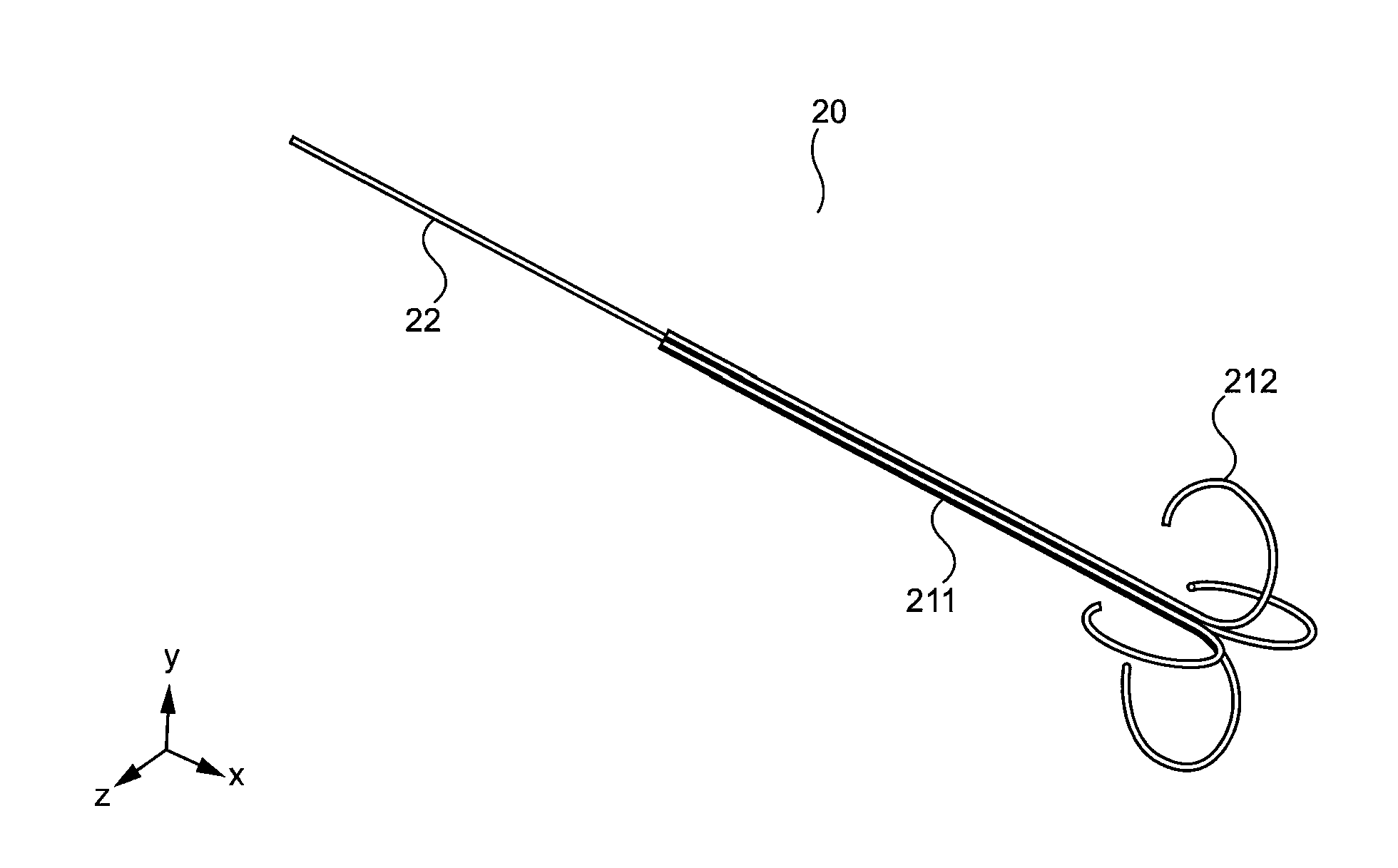
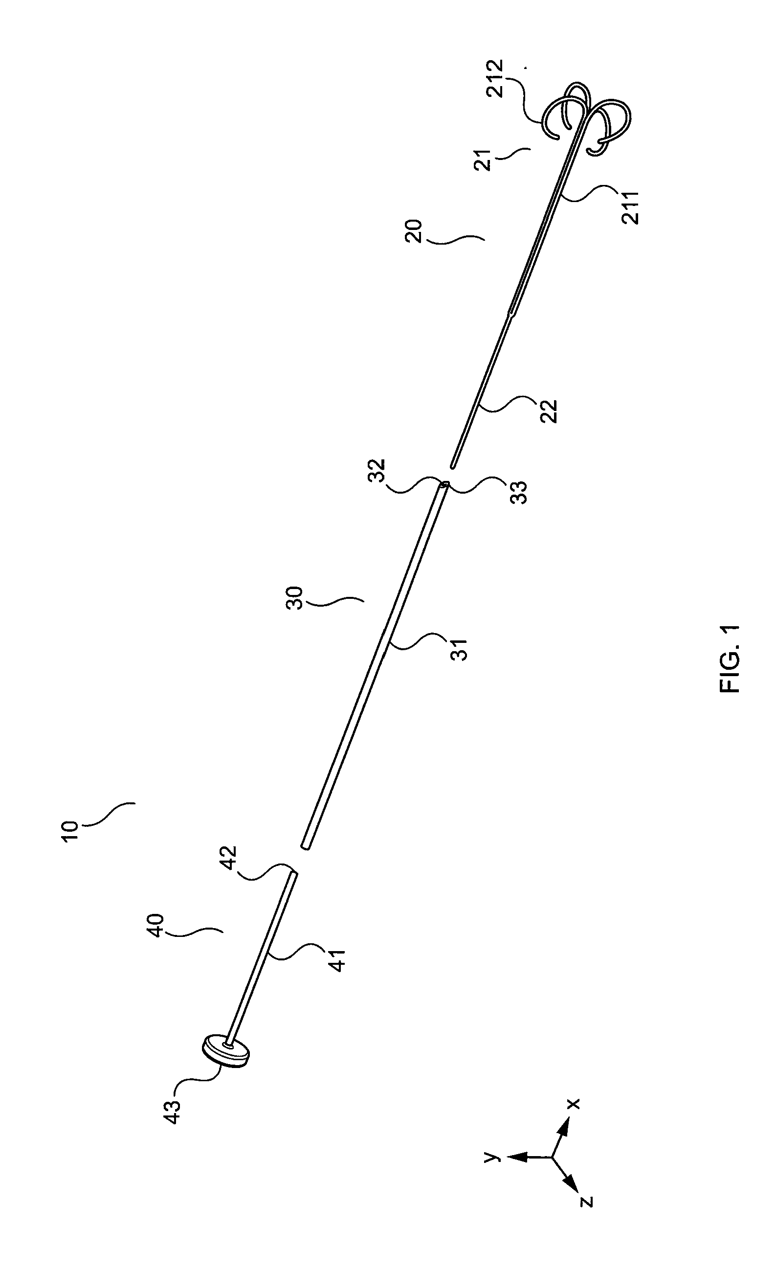
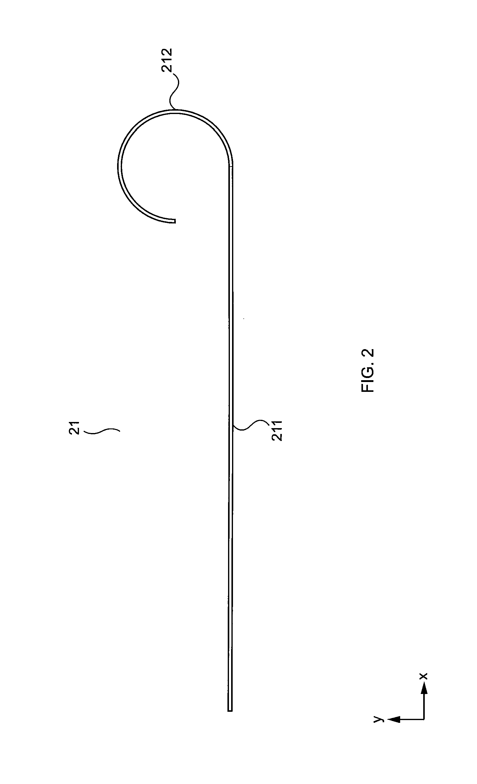
[0030]FIG. 1 is an exploded perspective view of a soft tissue mass excision guide system 10 including an encompassing device (sometimes referred to as a “soft tissue biopsy marking and encompassing device” or the “apparatus” or the “device”) according to an embodiment of the present invention. In preferred embodiments, the apparatus 10 includes a soft tissue marking and encompassing element (encompassing element) 20 comprising thre...


PUM


Abstract

The present invention is a device, system and method for surgical soft tissue lesion excision. The device is designed to be placed into a soft tissue lesion preferably under radiologic guidance and deployed. Once deployed, the shape memory wires of the soft tissue encompassing element form a wire cage surrounding the lesion which acts as an excision guide, encompassing and protecting the lesion during surgical excision of the lesion.

Description

[0001]This application claims the benefit of U.S. Provisional Application No. 61 / 869,825, filed Aug. 26, 2013, entitled SOFT TISSUE LESION EXCISION GUIDE, the disclosure of which is herein incorporated by reference.BACKGROUND OF THE INVENTION[0002]1. Field of the Invention[0003]The present invention relates generally to a device, system and method for performing surgical soft tissue lesion excision, more specifically, to a device, system and method for surgical soft tissue lesion excision performed after radiologic localization of the tissue lesion to be excised.[0004]2. Description of the Related Art[0005]In current practice, a patient with a radiologically identified lesion, for example, a breast lesion, which cannot be palpated by a physician, and thus cannot be surgically removed without some sort of guide or marker, is marked by placement of a radiologically guided wire which is placed into the tissue of the radiologically identified lesion. Subsequently, a surgeon during an op...

Claims


Application Information

Patent Timeline
Patent Type & Authority Applications(United States)
IPC IPC(8): A61B10/02A61B6/12
CPCA61B6/12A61B10/02A61B10/0233A61B2017/00867A61B90/39A61B2090/3908A61B2090/3937A61B2090/3987
Inventor DENNIS, TERAFRIEDLANDER, JACK
Owner EXCISURE LLC
Why Eureka
Try Eureka