

Whole cell assays and methods
a whole cell and assay technology, applied in the field of whole cell assays and methods, can solve the problem that the application of the method has not yet been widely utilized, and achieve the effect of improving the accuracy and reproducibility of the assay
- Summary
- Abstract
- Description
- Claims
- Application Information
AI Technical Summary
Problems solved by technology
Method used
Image

Examples
example 1
Pathway Shutdown Tests Showing Differentiated Response of Two Patients to Two Drugs
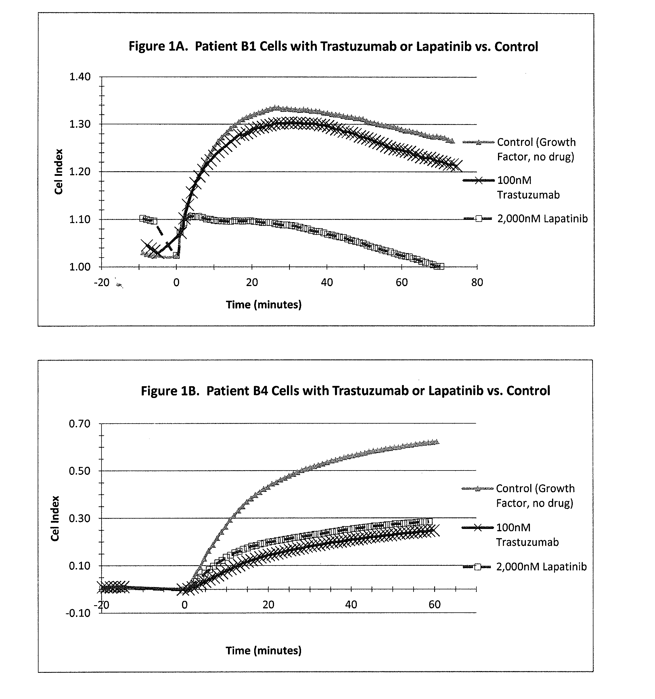
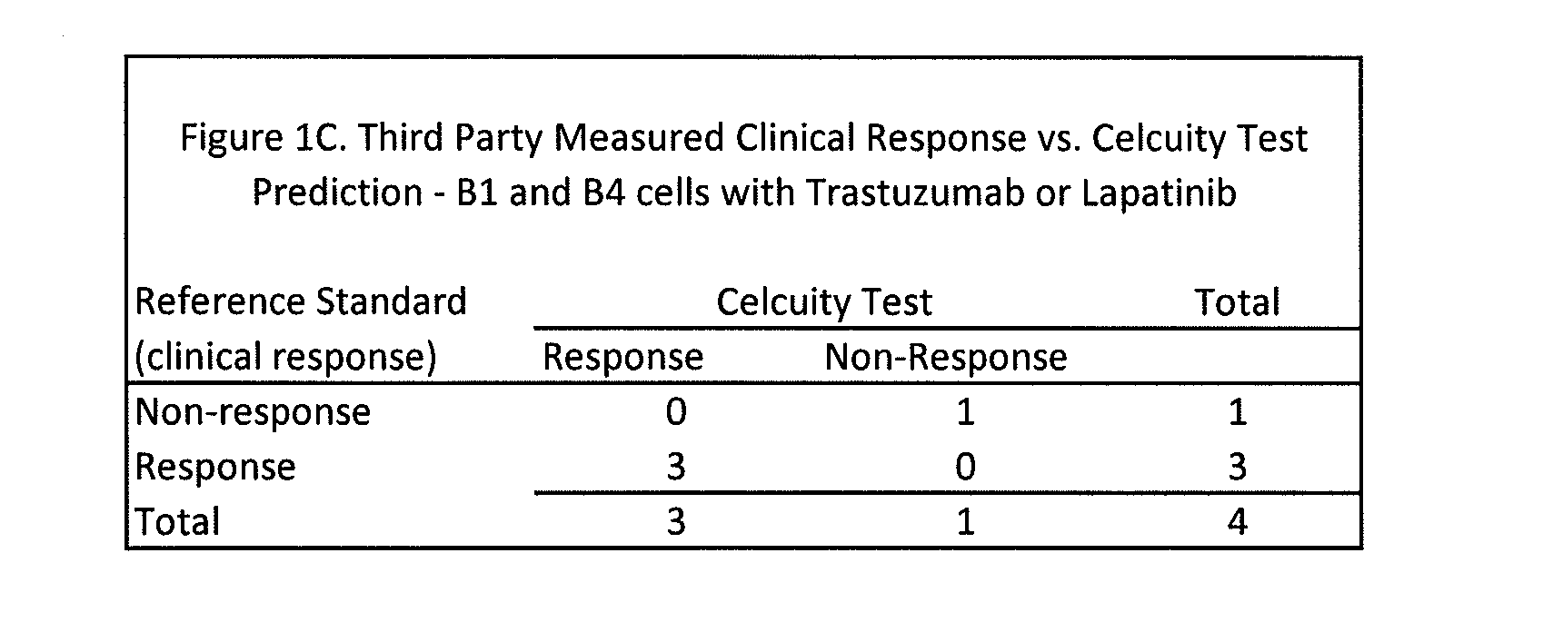
[0245]A CELx Pathway Shutdown test was performed using cells from two HER2 overexpressing breast cancer patients (Patient B1 and B4), two drugs (Lapatinib and Trastuzumab) that are indicated for HER2 positive breast cancers, and human epidermal growth factor (EGF). The physiologic change of the B1 and B4 cells during the test was measured with an impedance biosensor CReMS and the output from the CReMS is recorded in FIGS. 1A and 1B. The comparison of the CELx test results and the third party clinical reference is recorded in FIG. 1C. This example illustrates how the CELx test is able to predict the responsiveness that a patient will have to different targeted pathway drugs by using a CReMS to measure the physiological change in a patient's cells continuously over a period of several hours. This example also illustrates how the presence of a genetic biomarker, in this case an overexpressing HER2 gene, ...
example 2
Anti-Proliferative Tests Showing Differentiated Response of Two Patients to One Drug
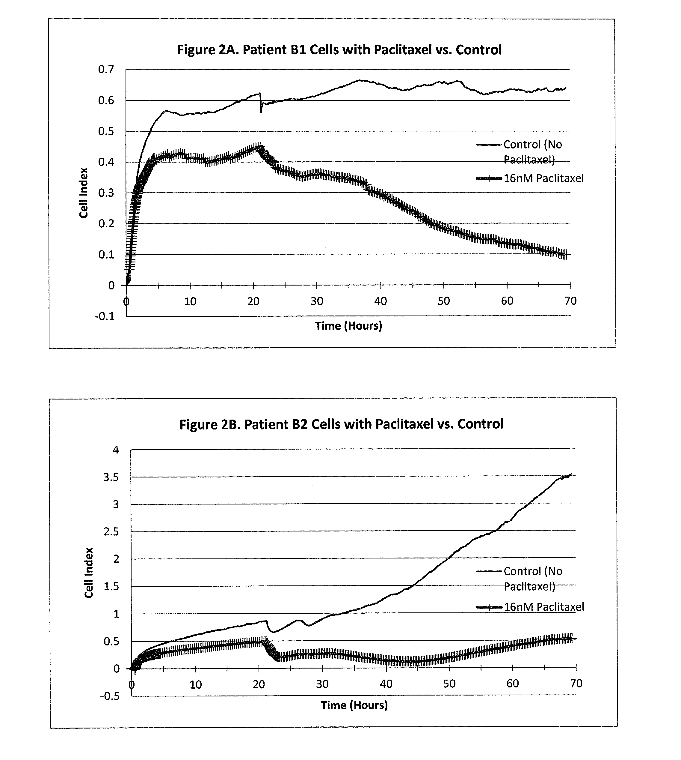
[0257]A CELx Anti-Proliferative test was performed using cells from two breast cancer patients (Patients B1 and B2) and the drug Paclitaxel. The physiologic change of the B1 and B2 cells during the test was measured with an impedance biosensor CReMS and the output from the CReMS is recorded in FIGS. 2A and 2B. The comparison of the CELx test results and the third party clinical reference is recorded in FIG. 2C. This example demonstrates the ability of the CELx test to predicting the efficacy of a therapeutic agent by measuring the physiologic change over the course of several days in a patient's cancer cells after an anti-proliferative drug is introduced. This example also demonstrates the role of a baseline, in this case, untreated patient cells, in measuring the results. In addition, the results recorded for patient B2 demonstrate the importance of monitoring the cells' physiological response on a ...
example 3
Combination Tests Showing Response of Two Patients to Two Drugs Taken Together
[0263]A CELx Combination test was performed using cells from two colon cancer patients (Patients C1 and C2), EGF, and a combination of two drugs indicated for colon cancer, Cetuximab and Irinotecan. The physiologic change of the C1 and C2 cells during the test was measured with an impedance biosensor CReMS and the output from the CReMS is recorded in FIGS. 3A and 3B. The comparison of the CELx test result and the third party clinical reference is recorded in FIG. 3C. This example demonstrates how the CELx test is able to predict the responsiveness that individual patients will have to a combination of two or more drugs in a way that cannot be done using genetic testing or expression profiling. The test also illustrates how the CELx test operates with colon cancer cells, in addition to breast cancer cells.
Materials and Methods
[0264]CReMS, microplate, reagents, and buffers: The CReMS, microplate, reagents, a...
PUM
| Property | Measurement | Unit |
|---|---|---|
| electrical frequency | aaaaa | aaaaa |
| adhesion | aaaaa | aaaaa |
| pH | aaaaa | aaaaa |
Abstract
Description
Claims
Application Information