Virtual endoscopic image display apparatus, method and program

a display apparatus and endoscopic technology, applied in tomography, applications, instruments, etc., can solve the problems of lowering the diagnostic efficiency, troublesome operation, and user trouble, and achieve the effect of convenient observation

Active Publication Date: 2015-10-08
FUJIFILM CORP
View PDF6 Cites 5 Cited by
  • Summary
  • Abstract
  • Description
  • Claims
  • Application Information

AI Technical Summary

Benefits of technology

The present invention relates to a virtual endoscopic image display program that allows a computer to generate and display virtual endoscopic images based on a three-dimensional image of a subject. The program can set a path for the virtual endoscope, receive specifications of points of interest, and obtain the distance of movement of the virtual endoscope from an initial viewpoint to a point of interest. The program can then generate and display virtual endoscopic images based on the distance of movement and the direction of view line vectors. The technical effect of this invention is that it provides a convenient and easy-to-use tool for generating and displaying virtual endoscopic images of a surrounding region of a point of interest.

Problems solved by technology

The patent text discusses a technique for extracting a three-dimensional image of a patient's tubular tissue, such as the colon, and using it for diagnosis purposes. The technical problem addressed in the text is how to accurately determine the path of an endoscope and generate a virtual endoscopic image to help navigate to a target point in the body.

Method used


Image

  • Virtual endoscopic image display apparatus, method and program
  • Virtual endoscopic image display apparatus, method and program
  • Virtual endoscopic image display apparatus, method and program

Examples


Embodiment Construction

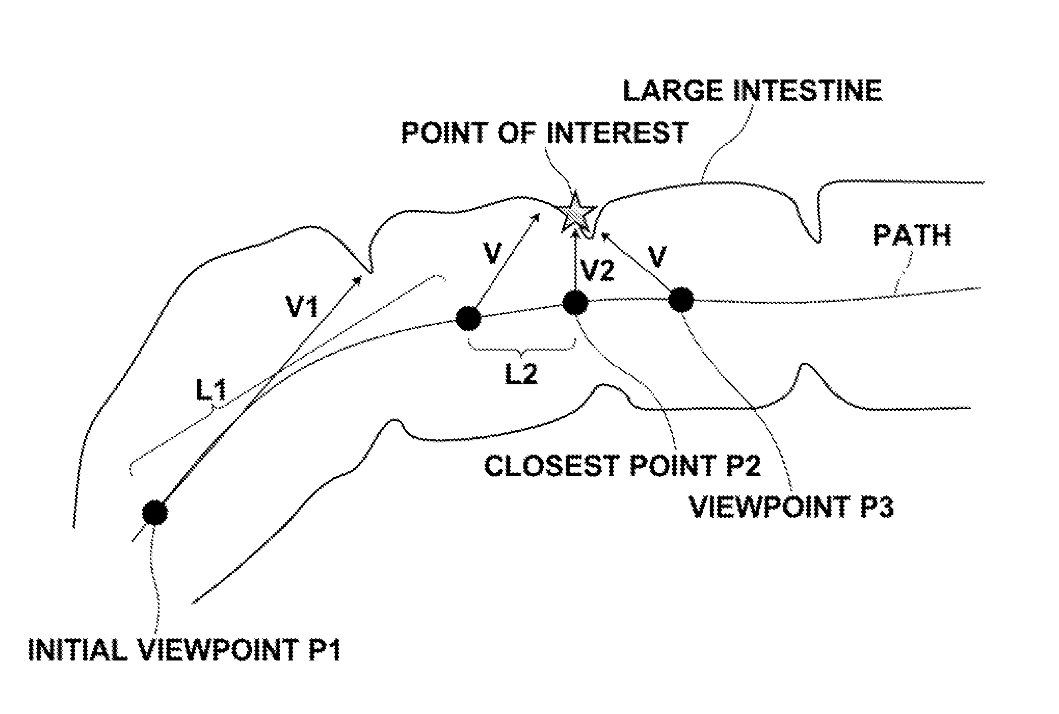
[0034]Hereinafter, an endoscopic image diagnosis support system using an embodiment of a virtual endoscopic image display apparatus, method and program of the present invention will be described in detail with reference to drawings. FIG. 1 is a schematic block diagram illustrating the configuration of an endoscopic image diagnosis support system according to an embodiment of the present invention.

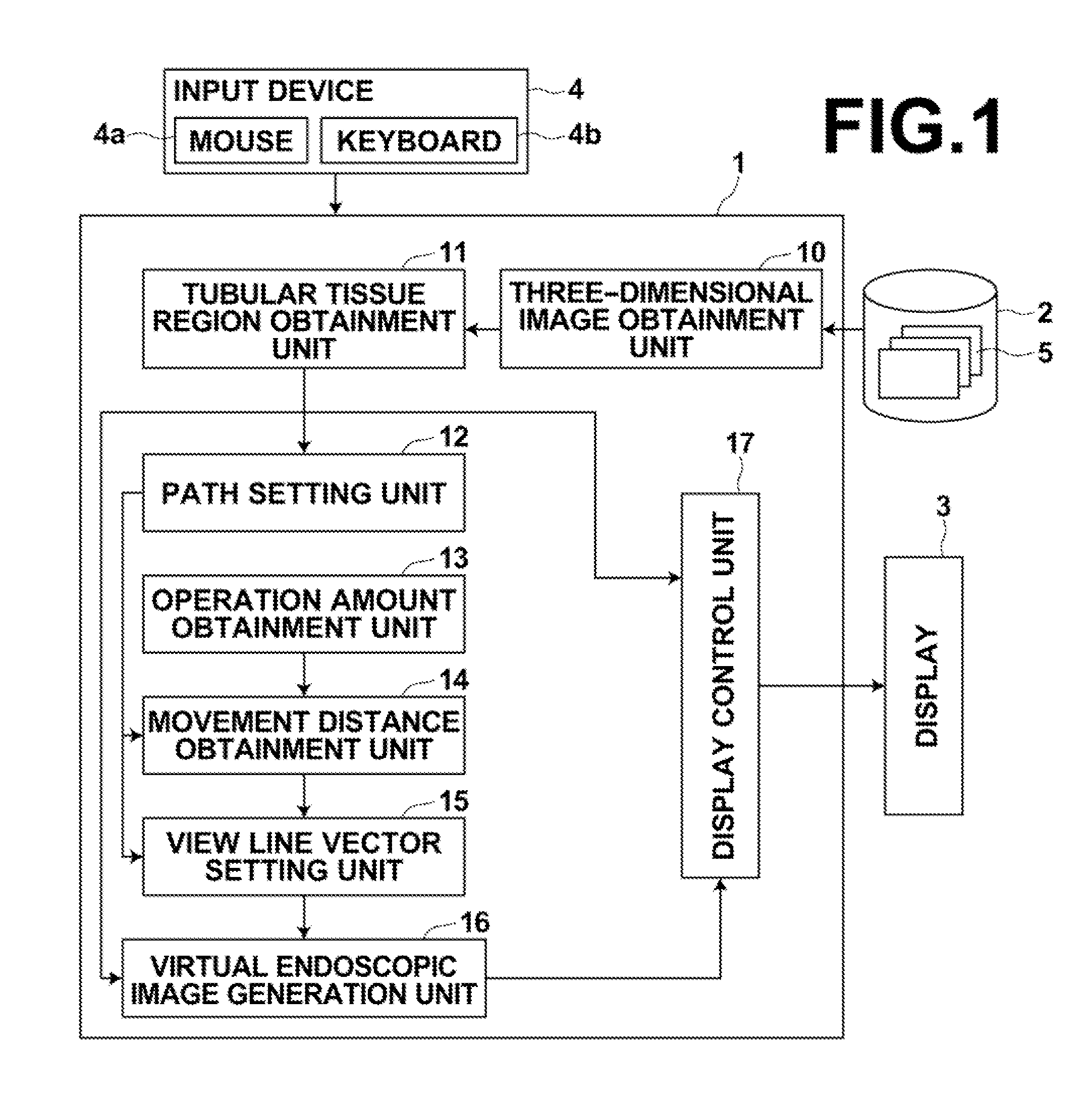
[0035]As illustrated in FIG. 1, the endoscopic image diagnosis support system according to an embodiment of the present invention includes an endoscopic image diagnosis support apparatus 1, a three-dimensional image storage server 2, a display 3 and an input device 4.

[0036]The endoscopic image diagnosis support apparatus 1 is a computer in which a virtual endoscopic image display program according to an embodiment of the present invention has been installed.

[0037]The endoscopic image diagnosis support apparatus 1 includes a central processing unit (CPU), a semiconductor memory and a storage...


PUM


Abstract

Specification of a point of interest is received. After then, a distance of movement on a path from an initial viewpoint is obtained based on an operation amount obtained at an operation unit, such as a mouse. View line vectors are set by sequentially changing the view line vectors from an initial view line vector with its start point located at the initial viewpoint to a shortest view line vector connecting a point of interest and a viewpoint at a shortest distance by moving, along the path, a viewpoint of a virtual endoscope on the path from the initial viewpoint closer to the point of interest and by changing the direction of a view line of the virtual endoscope closer to a direction toward the point of interest. Virtual endoscopic images are sequentially generated based on the set view line vectors, and the generated virtual endoscopic images are sequentially displayed.

Description


Claims


Application Information

Patent Timeline
Owner FUJIFILM CORP
Why Eureka
Try Eureka