Predictive Biomarkers for Detection of Organ Damage in Autoimmune Illnesses and Other Diseases

a technology of autoimmune illnesses and biomarkers, applied in the field of prediction biomarkers for organ damage detection in autoimmune illnesses and other diseases, to achieve the effect of preventing organ damag

Active Publication Date: 2017-02-02
FLORIDA INTERNATIONAL UNIVERSITY
View PDF0 Cites 5 Cited by
  • Summary
  • Abstract
  • Description
  • Claims
  • Application Information

AI Technical Summary

Problems solved by technology

Autoimmune diseases develop when the immune system misidentifies a self-antigen as a foreign antigen and produces destructive responses to the self-antigen.

Method used


Image

  • Predictive Biomarkers for Detection of Organ Damage in Autoimmune Illnesses and Other Diseases
  • Predictive Biomarkers for Detection of Organ Damage in Autoimmune Illnesses and Other Diseases
  • Predictive Biomarkers for Detection of Organ Damage in Autoimmune Illnesses and Other Diseases

Examples


example 1

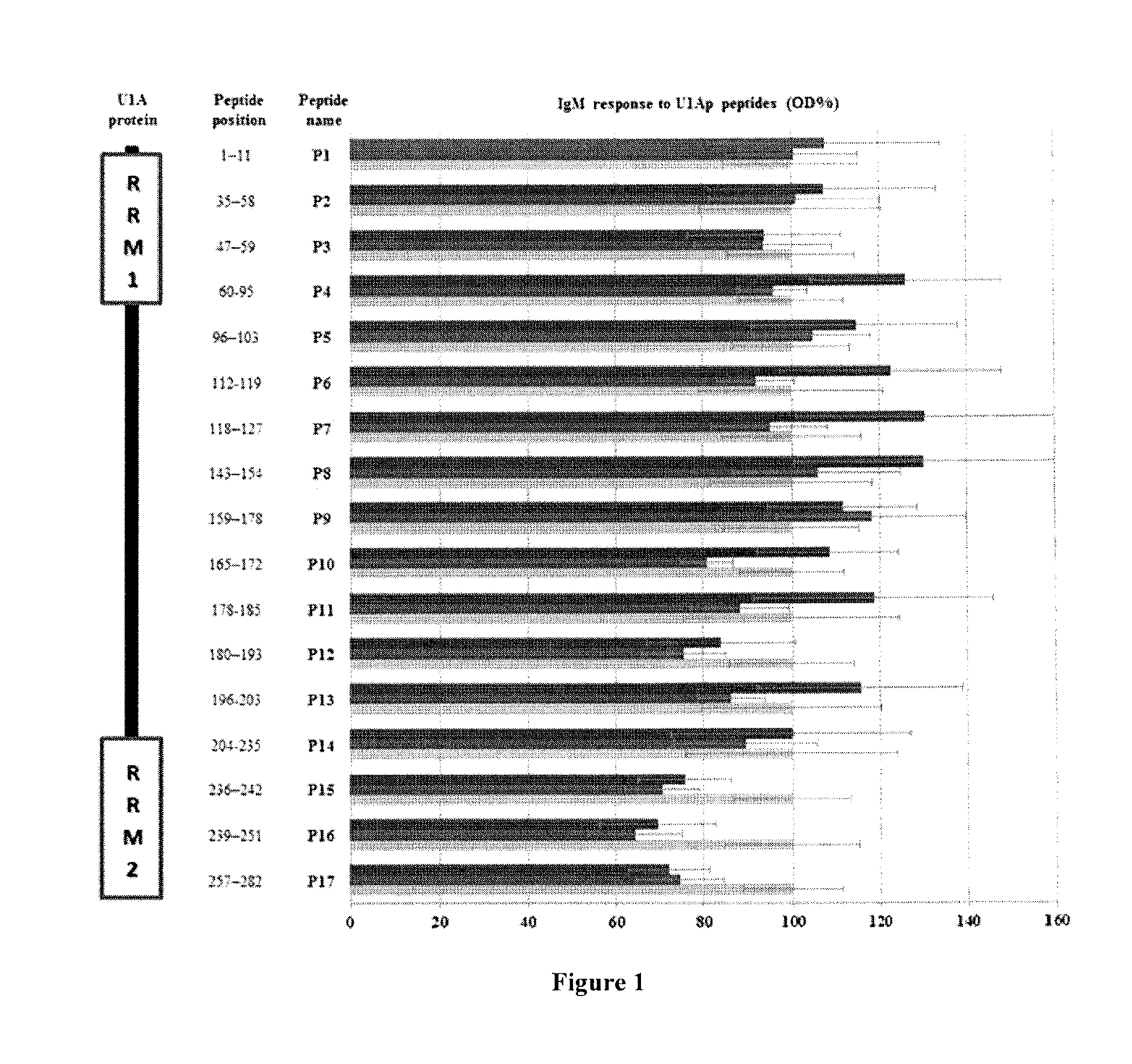
Auto-Antigenic IgM Responses to U1Ap

[0160]Indirect ELISAs were employed to assess IgM responses for U1Ap peptides in sera from SLE, MCTD and healthy individuals. In general, SLE patients exhibited significantly higher IgM anti-U1Ap titers when compared to the healthy individuals (P1-P11 and P13, FIG. 1). In contrast, except for P5, P8 and P9, MCTD patients had significantly lower IgM responses to U1Ap peptides than healthy individuals (FIG. 1). Interesting, anti-U1Ap-P16 IgM reactivity was significantly lower in both SLE and MCTD patients compared to healthy controls (p≦0.04) (FIG. 1). SLE patients showed trends toward significantly higher IgM reactivity for U1Ap subunits than MCTD patients (P4, P6, P7, P10, P11 and P13 were the most antigenic U1Ap fragments against IgM with 1.3 fold significantly higher levels in SLE than MCTD patients), but the differences were not significant (p>0.05, FIG. 1). Specifically, P7 and P17 represented the biggest (137%) and smallest (96%) differences ...

example 2

Auto-Antigenic IgM Response is Directed to Non-RNA Recognition Domain Regions of U1Ap

[0162]To compare the IgM reactivity in RNA recognition domain versus non-RNA recognition domain fragments of U1Ap, the average peptide reactivity corresponding to regions identified as RNA recognition motif (RRM) 1 (P1-P4), RRM2 (P14-17) and the non-domain area (P4-P13) were calculated in SLE, MCTD and healthy populations. Patients showed elevated IgM anti-U1Ap responses to non-domain areas of the U1Ap (P5-P13) when compared to RRM1 (P1-P4) and RRM2 (P14-P17) (FIG. 1). However, only the SLE and not the MCTD patient subgroup exhibited significantly elevated average IgM response to peptides in U1Ap non-domain regions when compared to the average IgM reactivity for peptides covering RRM1 or RRM2 (p≦0.003). The IgM response to the U1Ap C-terminal end, which encompasses RMM2, was noticeably significantly lower in both SLE and MCTD patients than in control samples (FIG. 1).

example 3

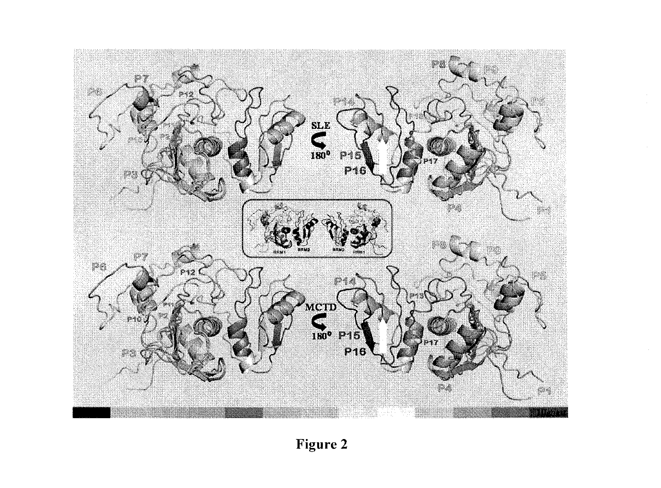
IgM Derived U1Ap 3D Epitope Maps for SLE and MCTD Patient

[0163]The IgM reactivity for each U1Ap peptide monitored in sera from SLE and MCTD patients was used to create two independent epitope maps of U1Ap (FIG. 2). In general, the IgM reactivity was not always directed to exposed fragments of the U1Ap, given that superficial peptides appeared to have equal chance to be highly antigenic (P7 in SLE and P9 in MCTD) or not (P17 in SLE and P4 in MCTD) (FIG. 2). Likewise, different molecular structures like α-helices (P4), β-sheets (P15) and loops (P8) seemed to elicit antigenic IgM responses to U1Ap. However, β-sheets were the least antigenic of all three forms (P15 and P16 in FIG. 2). When comparing the SLE and MCTD subgroups, significantly higher U1Ap IgM reactivity was directed to superficial peptides with α-helical structure in both SLE (P7) and MCTD (P4) (FIG. 2). The lowest IgM responses were observed for peptides corresponding to exposed β-sheet fragments of the U1Ap in both SLE a...


PUM

PropertyMeasurementUnit
pressureaaaaaaaaaa
IgM reactivityaaaaaaaaaa
optical density percentageaaaaaaaaaa

Abstract

The invention provides methods for identifying the presence of, or an increased risk of developing, organ damage in a subject having an autoimmune disease, for example, Systemic Lupus Erythematosus (SLE) or Mixed Connective Tissue Disease (MCTD), or other disease in which the lungs and / or kidneys are involved. In one embodiment, a significantly increased combined IgM reactivity against the peptides having the sequences of SEQ ID NOs: 4, 9, 12, and 15 in a sample obtained from a patient compared to a healthy control indicates lung damage or an increased risk of developing lung damage in the subject. In another embodiment, a significantly increased combined IgM reactivity against the peptides having the sequences of SEQ ID NOs: 2, 8, 12, 14, 15, and 17, in a sample from a patient compared to a healthy control indicates kidney damage or an increased risk of developing kidney damage in the subject.

Description

CROSS REFERENCE TO RELATED APPLICATION(S)[0001]This application claims the benefit of U.S. Provisional Application Ser. No. 62 / 199,118, filed Jul. 30, 2015, which is hereby incorporated by reference in its entirety, including any figures, tables, or drawings.GOVERNMENT SUPPORT[0002]The subject invention was made with government support under a research project supported by National Institute of Health under Grant Nos. 2R25 GM061347-13 and AR48805, and S06-GM08205. The government has certain rights in this invention.[0003]The Sequence Listing for this application is labeled “SeqList-25Jul16.txt”, which was created on Jul. 25, 2016, and is 5 KB. The entire content is incorporated herein by reference in its entirety.BACKGROUND OF INVENTION[0004]Autoimmune diseases develop when the immune system misidentifies a self-antigen as a foreign antigen and produces destructive responses to the self-antigen.[0005]Patients with autoimmune disease frequently develop kidney involvement and / or lung ...

Claims


Application Information

Patent Timeline
Patent Type & Authority Applications(United States)
IPC IPC(8): G01N33/564
CPCG01N33/564G01N2800/50G01N2800/104G01N33/6854
Inventor MESA, ANNIASOMARELLI, JASON A.
Owner FLORIDA INTERNATIONAL UNIVERSITY
Why Eureka
Try Eureka