Indication-Dependent Display of a Medical Image

Active Publication Date: 2017-03-23
BRAINLAB
View PDF1 Cites 8 Cited by
  • Summary
  • Abstract
  • Description
  • Claims
  • Application Information

AI Technical Summary

Benefits of technology

[0038]The atlas data is in particular multi-modal atlas data describing an image-based model of at least part of the human body comprising the anatomical structure. Multi-modal atlas data is atlas data which contains image information about anatomical structures which has been generated by application of different imaging modalities. For example, the atlas data may contain an image-based model which has been generated by computed x-ray tomography and an image-based model which has been generated by magnetic resonance tomography. This allows receiving different image information for generating the image-based model. For ex

Problems solved by technology

When assessing digital medical images in particular in three dimensions (such as computed x-ray tomographies or magnetic resonance tomographies), it may however b

Method used


Image

  • Indication-Dependent Display of a Medical Image
  • Indication-Dependent Display of a Medical Image
  • Indication-Dependent Display of a Medical Image

Examples


Example

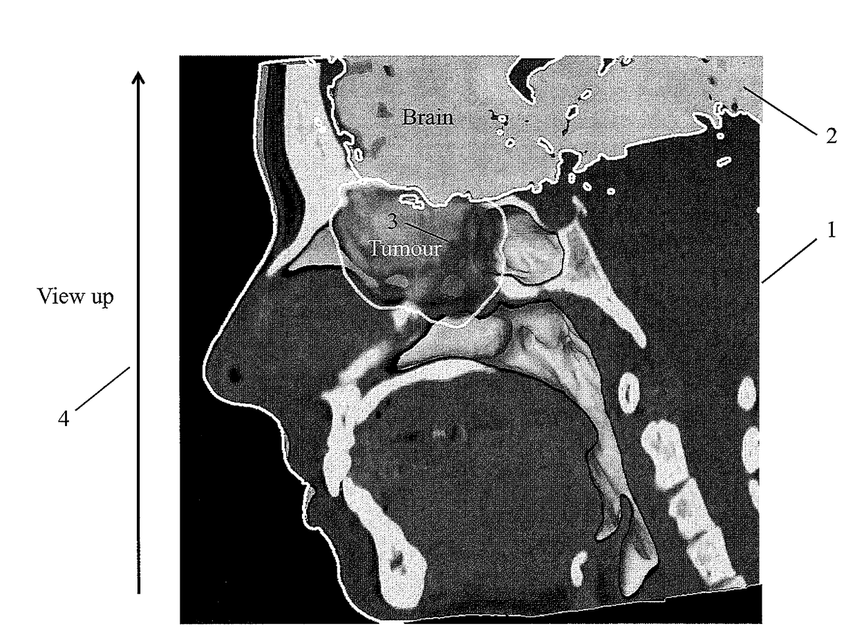
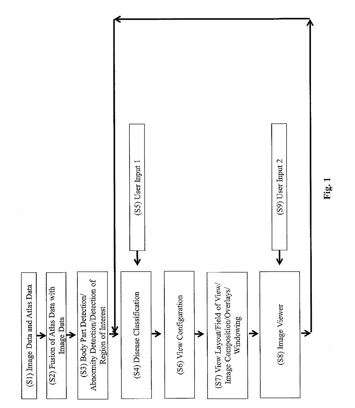
[0065]FIG. 1 shows the flow of steps of the method in accordance with the invention according to a preferred embodiment. The invention is not to be construed as being limited to the steps shown in FIG. 1.

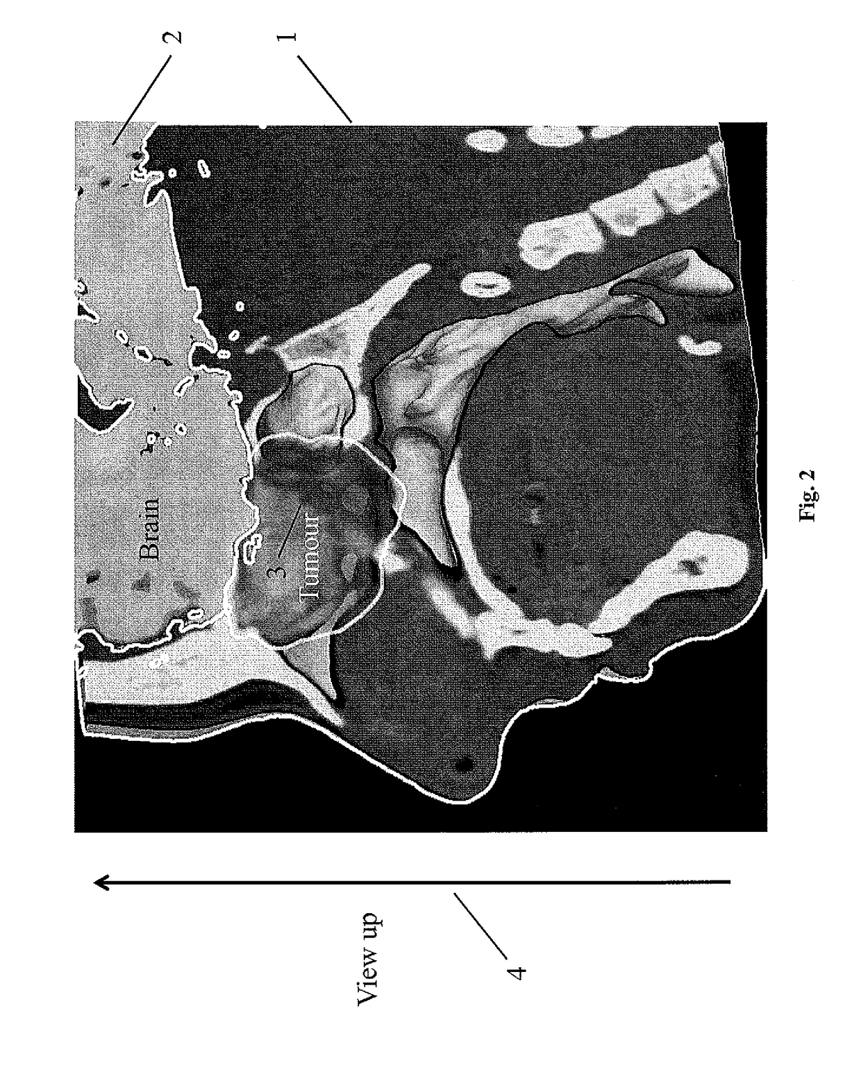
[0066]In step S1, image data representing the patient medical image data is acquired along with the atlas data. In step S2, the atlas data is fused to the image data in order to determine the atlas-patient transformation data. On the basis of the thus-acquired patient image data and atlas data, the region of interest within the patient image data is detected in step S3 which encompasses in particular also the determination of the atlas-patient transformation data. Preferably, the detection of the region of interest of the patient image data is a body part detection, more preferably abnormality detection, i.e. detection of anatomic abnormalities such as a pathologic region (for example a tumour or a bone fracture) in the patient image data as a result of the fusion conducted in step ...


PUM


Abstract

The present invention relates to a medical data processing method of determining an image of an anatomical structure of a patient's body, the method comprising the following steps which are constituted to be executed by a computer: a) acquiring (S1) GP atlas data describing an image-based model of at least part of a human body comprising the anatomical structure; b) acquiring (S1) patient medical image data describing a patient-specific medical image of the anatomical structure in the patient's body, wherein the patient medical image data comprises in particular three-dimensional image information c) determining (S2), based on the atlas data and the patient medical image data, atlas-patient transformation data describing a transformation between the image-based model and the anatomical structure in the patient's body; d) acquiring (S3, S4) medical indication data describing a medical indication which the anatomical structure is subject to; e) acquiring (S7) imaging parameter data describing at least one imaging parameter for generating, from the image-based model, an image of the anatomical structure in dependence on the medical indication data: f) determining (S8) indication image data describing an indication-specific image (1) of the anatomical structure in the patient, wherein the indication image data is determined (S8) based on the patient medical image data and the atlas-patient transformation data and the medical indication data and the imaging parameter data.

Description

[0001]The present invention is directed to a data processing method of determining an image of an anatomical structure of a patient's body in dependence on a medical indication.[0002]When planning a medical procedure such as a therapeutic or surgical procedure, medical personnel such as a physician commonly wishes to have an image of the anatomical structure which shall undergo the medical procedure in order to have a visual impression of his working area. In many cases, the person planning the medical procedure wishes to have the anatomical structure presented in a view which is useful for conducting the planning. Such a view may for example be a standard view which in the medical arts is known to represent a view having the most useful information content for planning the procedure. For example, in the case of radiotherapy or radiosurgery on a tumour, the anatomical structure in which the tumour is located should be displayed in the image such that the tumour is visible. As anothe...

Claims


Application Information

Patent Timeline
IPC IPC(8): G06T7/00G06T11/60
CPCG06T7/0012G06T2207/10004G06T11/60G06T19/20
Inventor VILSMEIER, STEFAN
Owner BRAINLAB
Why Eureka
Try Eureka