Method and system for automated analysis of cell images

a cell image and automated analysis technology, applied in image analysis, image enhancement, instruments, etc., can solve the problem of not providing a solution for different clustering types of cells

Inactive Publication Date: 2017-03-30
KONICA MINOLTA LAB U S A INC
View PDF7 Cites 17 Cited by
  • Summary
  • Abstract
  • Description
  • Claims
  • Application Information

AI Technical Summary

Benefits of technology

[0004]In consideration of the above issues, it would be desirable to have a system and method for the cell segmentation, for example, of microscopy cell images by fi

Problems solved by technology

Although there are methods and systems, which perform cell segmentation, the

Method used


Image

  • Method and system for automated analysis of cell images
  • Method and system for automated analysis of cell images
  • Method and system for automated analysis of cell images

Examples


Embodiment Construction

[0030]Reference will now be made in detail to the present preferred embodiments of the invention, examples of which are illustrated in the accompanying drawings. Wherever possible, the same reference numbers are used in the drawings and the description to refer to the same or like parts.

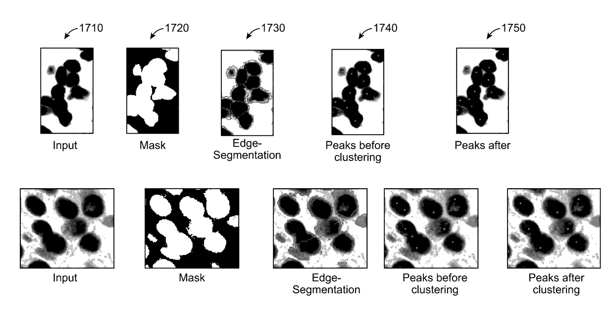
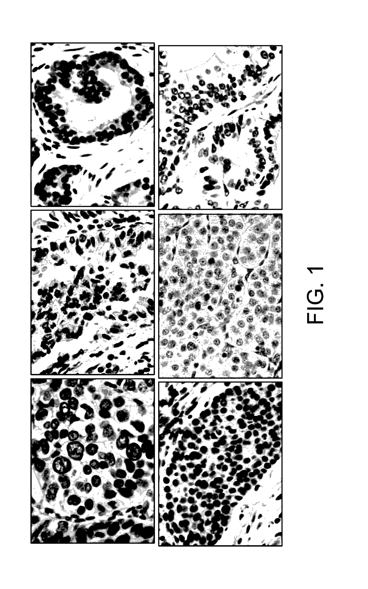
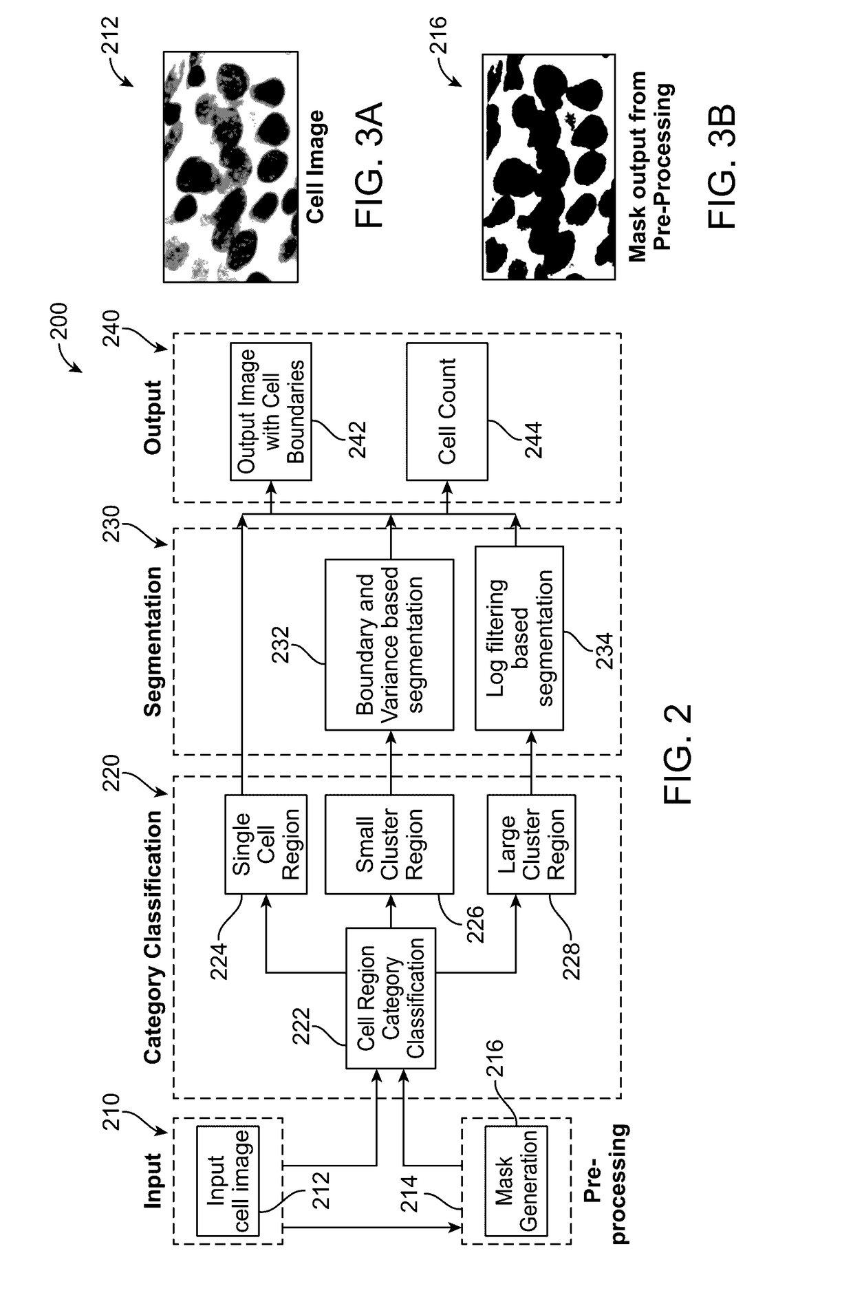
[0031]In accordance with an exemplary embodiment, unlike many other methods, a system and method are disclosed, which can be suitable for different sizes of cells present in a single image, for example, irrespective of the cell size (small or large), and which can extract the cell boundaries. FIG. 1 illustrates various kinds of cell images, which can be analyzed and processed in accordance with the systems and methods as disclosed herein.

[0032]FIG. 2 shows a block diagram for a system 200 for cell segmentation in accordance with an exemplary embodiment. As shown in FIG. 2, the system 200 can include an input module 210, a pre-processing module 214, a category classification module 220, a segmentation...


PUM


Abstract

A method, a computer readable medium, and a system are disclosed for cell segmentation. The method including generating a binary mask from an input image of a plurality of cells, wherein the binary mask separates foreground cells from a background; classifying each of the cell regions of the binary mask into single cell regions, small cluster regions, and large cluster regions; performing, on each of the small cluster regions, a segmentation based on a contour shape of the small cluster region; performing, on each of the large cluster regions, a segmentation based on a texture in the large cluster regions; and outputting an image with cell boundaries.

Description

CROSS-REFERENCES TO RELATED APPLICATIONS[0001]This application claims priority to U.S. Provisional Patent Application Ser. No. 62 / 235,076, filed on Sep. 30, 2015, the entire content of which is incorporated herein by reference.FIELD OF THE INVENTION[0002]The present disclosure relates to a method and system for automated analysis of cell images, and more particularly for a method and system for automated cell segmentation for microscopic cell images, which can be categorized into single cells, small clusters, and large clusters, and wherein cell boundaries can be extracted from the cell images.BACKGROUND OF THE INVENTION[0003]In the biomedical imaging domain, segmenting the touching cell nuclei can be a very important step in image analysis. Although there are methods and systems, which perform cell segmentation, these systems do not provide a solution for different clustering types of cells.SUMMARY OF THE INVENTION[0004]In consideration of the above issues, it would be desirable to...

Claims


Application Information

Patent Timeline
IPC IPC(8): G06T7/00G06K9/52G06K9/62G06T7/60G06K9/66G06K9/46
CPCG06T7/0081G06K9/66G06T7/0085G06K9/4604G06K9/6267G06T2207/20144G06K9/52G06T7/0002G06K9/4671G06T7/0042G06K2009/4666G06T7/60G06T2207/30024G06T7/73G06T7/11G06T7/13G06T7/194G06F18/24
Inventor PARADKAR, FORAM MANISHZHANG, YONGMIANZHU, JINGWENGU, HAISONG
Owner KONICA MINOLTA LAB U S A INC
Why Eureka
Try Eureka