

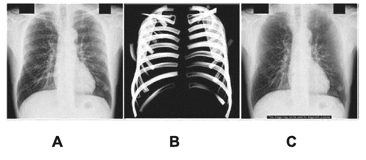
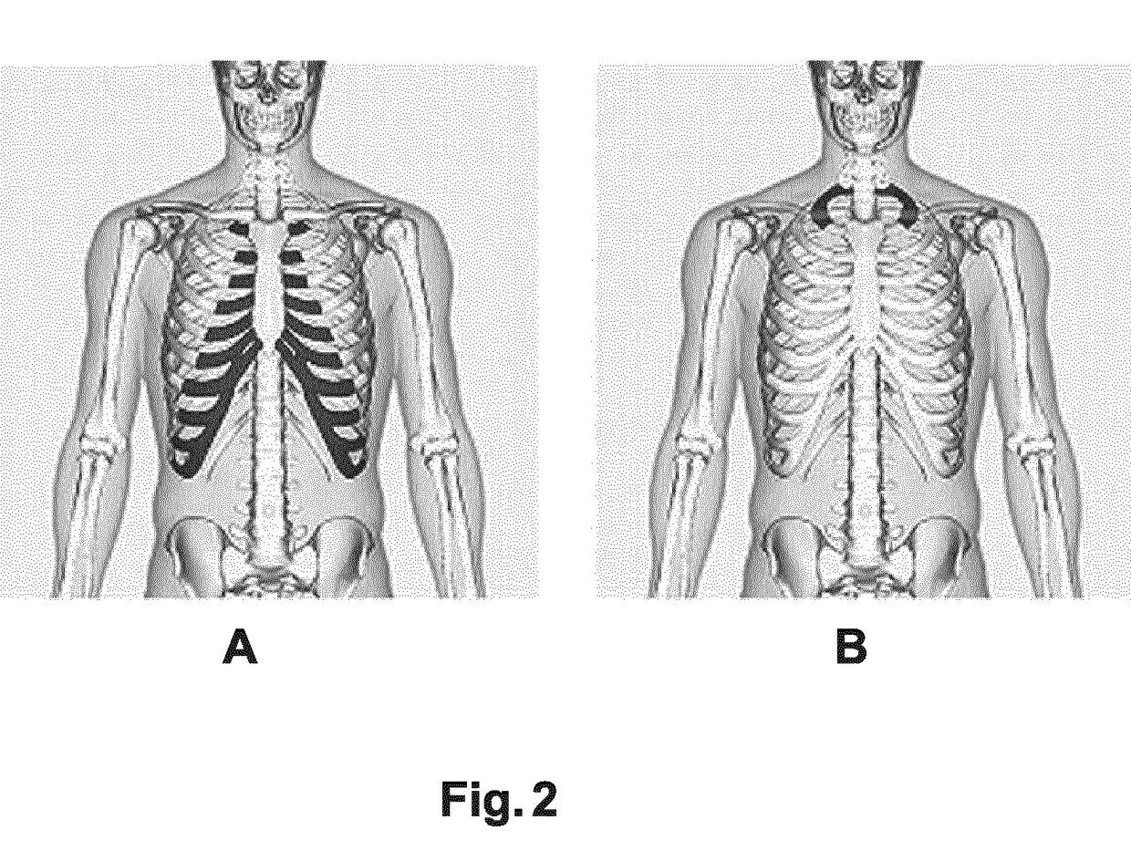
Silhouette display for visual assessment of calcified rib-cartilage joints
a rib-cartilage joint and image processing technology, applied in the field of image processing, can solve the problems of affecting the interpretation of image information, and affecting the interpretation of radiographic images,
- Summary
- Abstract
- Description
- Claims
- Application Information
AI Technical Summary
Benefits of technology
Problems solved by technology
Method used
Image

Examples
Embodiment Construction
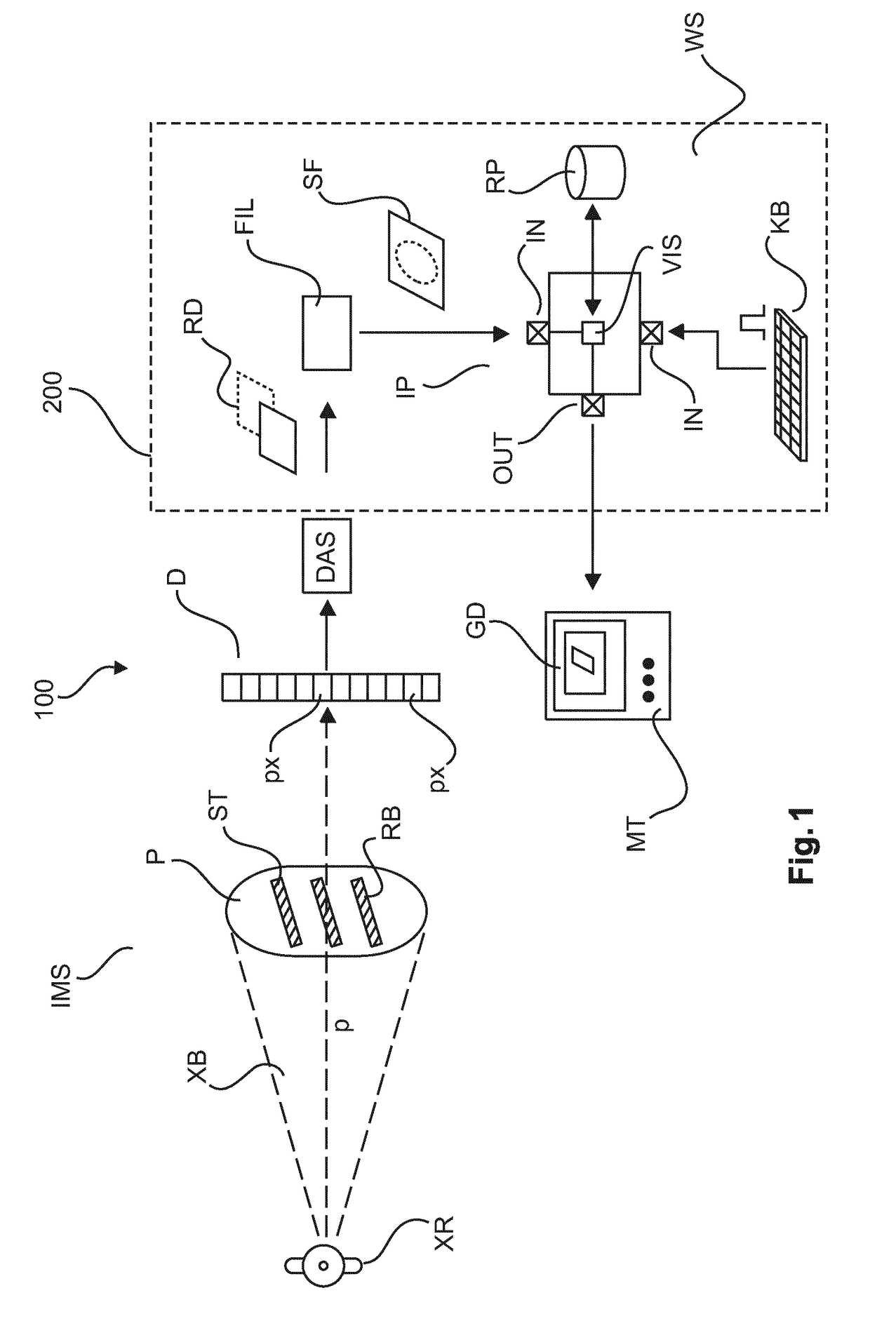
[0033]With reference to FIG. 1 there is shown an arrangement 100 including an acquisition component IMS for acquisition of radiographs RD (X-ray images) of an object P and a system 200 to support visual analysis of the radiographs so acquired.
[0034]The image acquisition component IMS includes an X-ray source XR and a radiation sensitive detector D. The x-ray source XR and the detector D are suitably supported in a mechanical structure such as a gantry (not shown). The image acquisition component IMS may include but is not limited to an x-ray imager of the C-arm type or may include a CT image apparatus or any other apparatus suitable for the acquisition of x-ray based radiographs.
[0035]The x-ray source XR is configured to emit, during an imaging session, x-ray radiation. More particularly, X-ray beam XB passes through an examination region and then impinges on a radiation sensitive surface of the detector D. The radiation sensitive surface is made up from one or more rows of detector...
PUM
 Abstract
Description
Claims
Application Information