
Identification and isolation of neural stem cells and neurosphere initiating cells

a neural stem cell and initiating cell technology, applied in the field of neurology and developmental biology, can solve the problems of hampered efforts to assess their properties, inability to directly study qnscs or nics as they exist in vivo, and uncertainty regarding the relationship between qnscs and nics

Inactive Publication Date: 2017-07-06
BOARD OF RGT THE UNIV OF TEXAS SYST
View PDF0 Cites 4 Cited by
  • Summary
  • Abstract
  • Description
  • Claims
  • Application Information

AI Technical Summary

Benefits of technology

The present patent provides a method for isolating a population of neural stem cells from adult mouse brain that exhibit specific expression patterns of Glast, EGFR, PlexinB2, CD24, O4, PSA-NCAM, Ter119, and CD45. These cells can be subventricular zone (SVZ) cells, which are known to form neurospheres and differentiate into various cell types in culture. The isolated cell population can also be enriched for cells expressing GFAP+ and cells recombined by GFAP-CreERT2. The isolated cells can be cultured and treated with temozolomide (TMZ), a drug used to treat glioblastoma. The patent also provides a kit containing antibodies against the above markers for use in identifying and isolating the neural stem cells.

Problems solved by technology

However, in the adult forebrain it remains uncertain whether these colonies are formed by quiescent neural stem cells (qNSCs) or by mitotically active and shorter-lived multipotent progenitors (Morshead et al., 1994; Doetsch et al., 2002; Reynolds and Rietze, 2005; Pastrana et al., 2009; Pastrana et al., 2011).
Moreover, reliance upon retrospective colony-formation assays makes it impossible to directly study qNSCs or NICs as they exist in vivo.
These studies have provided a critical framework for understanding the SVZ neurogenic lineage, though the inability to purify live cells from each stage of this hierarchy has hampered efforts to assess their properties.
The inability to prospectively identify and isolate uncultured stem cells from the central nervous system (CNS) has contributed to uncertainty regarding the relationship between qNSCs and NICs.
Efforts toward prospective identification have therefore generated conflicting results about whether NICs are quiescent or mitotically active in vivo and regarding their relationship to NSCs.
However, an important question that they have not been able to address directly is whether those genes are necessary for NSC maintenance in vivo.
Thus, it has not been possible to test whether Bmi-1 is autonomously required by NSCs in the adult brain or whether NSCs differ from NICs in their dependence upon Bmi-1.

Method used


Image

  • Identification and isolation of neural stem cells and neurosphere initiating cells
  • Identification and isolation of neural stem cells and neurosphere initiating cells
  • Identification and isolation of neural stem cells and neurosphere initiating cells

Examples


example 1

Materials and Methods

[0095]Mice. C57B1 / 6 mice were maintained in standard cages with water and standard diet (Teklad 2916) ad libitum. Rosa26CAG-loxp-Stop-loxp-tdTomato(Ai14) (referred to here as loxp-tdTomato) (Madisen et al., 2010), Gli1CreERT2 (Ahn and Joyner, 2005), Sox2CreERT2 (Arnold et al., 2011), Dlx1CreERT2 (Taniguchi et al., 2011), Tg(Glast-CreERT) (Wang et al., 2012), Tg(Nestin-Cre) (Tronche et al., 1999) and Tg(Ubc-GFP) (Schaefer et al., 2001) mice were obtained from The Jackson Laboratory. Tg(GFAP-CreERT2) (Hirrlinger et al., 2006) mice were provided by Frank Kirchhoff. Tg(Nestin-GFP) (Birbrair et al., 2011) and Tg(Nestin-mCherry) (Ding et al., 2012) mice were kindly provided by Grigori Enikopalov. Tg(Nestin-CreERT2) mice were provided by G. Fishell (Balordi and Fishell, 2007). All mice were backcrossed onto a C57BL / Ka background for at least 3 generations prior to analysis. For BrdU pulses up to 24 hours, 100 mg of BrdU / kg body mass dissolved in PBS was injected i.p. e...

example 2

Results

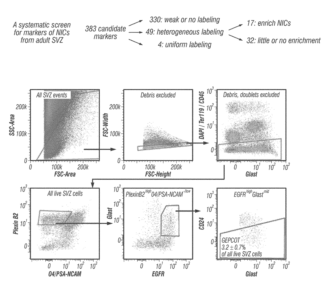
[0106]Prospective identification of NICs. The inventors enzymatically dissociated adult mouse SVZ cells then sorted cells by flow cytometry into non-adherent cultures at clonal density (0.66 cells / μl of culture medium). The inventors always replated neurospheres to adherent secondary cultures to assess differentiation into TuJ1+ neurons, GFAP+ astrocytes, and O4+ oligodendrocytes. On average, 1.8±0.4% of SVZ cells formed neurospheres (>50 μm diameter) and 75% of those neurospheres underwent multilineage differentiation (1.4±0.3% of SVZ cells).

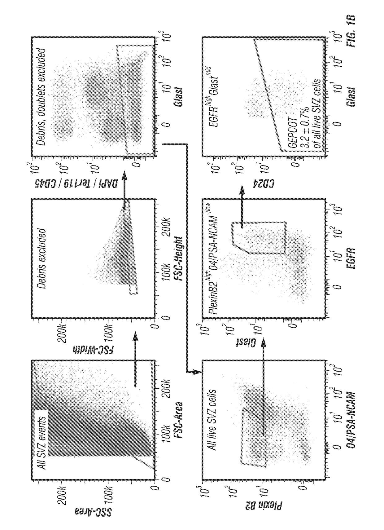
[0107]The inventors systematically screened 383 antibodies against 330 distinct cell surface antigens (data not shown) to identify markers that could enrich NICs (FIG. 1A). The inventors identified 49 markers by flow cytometry that were heterogeneously expressed among dissociated SVZ cells. For each of these markers they sorted SVZ cells that differed in their level of staining into non-adherent cultures and assessed neurosphere formatio...

example 3

Discussion

[0142]By screening almost four hundred antibodies against distinct cell surface antigens, the inventors identified two phenotypically and functionally distinct populations of neural stem / progenitor cells from the adult mouse SVZ. GEPCOT cells were highly enriched for NICs (FIG. 1C) and highly mitotically active in vivo (FIG. 1D) but persisted only transiently in the SVZ based on fate mapping with Ascl1CreERT2 or Dlx1CreErT2 (FIGS. 2B-C). In contrast, pre-GEPCOT cells lacked the ability to form neurospheres or adherent colonies in culture (FIG. 4B), and were quiescent in vivo (FIG. 4C) but were long-lived in the SVZ based on fate mapping with the stem cell markers Glast-CreERT (Wang et al., 2012), GFAP-CreERT2 (Giachino et al., 2013), Sox2CreERT2 (Arnold et al., 2011), and Gli1CreERT2 (Ahn and Joyner, 2005; Lee et al., 2012) (FIGS. 4F-J). In contrast to GEPCOT NICs, pre-GEPCOT cells were resistant to TMZ (FIGS. 5E-F). Although TMZ eliminated virtually all NICs from the SVZ ...


PUM

PropertyMeasurementUnit
depthaaaaaaaaaa
frequencyaaaaaaaaaa
body massaaaaaaaaaa

Abstract

The disclosure reports on the identification and isolation of adult mouse lateral ventricle subventricular zone (SVZ) neurosphere initiating cells (NICs) by flow cytometry on the basis of GlastmidEGFRhighPlexinB2highCD24− / lowO4 / PSA-NCAM− / lowTer-119 / CD45− markers (GEPCOT cells). These cells are highly mitotic and short-lived in vivo based on fate-mapping with Ascl1CreERT2 and Dlx1CreERT2. In contrast, pre-GEPCOT cells were quiescent, expressed higher Glast, and lower EGFR and PlexinB2. Pre-GEP-COT cells could not form neurospheres but expressed the stem cell markers Glast-CreERT, GFAP-CreERT2, Sox2CreERT2, and Gli1C-reERT2 and were long-lived in vivo. While GEPCOT NICs were ablated by temozolomide, pre-GEPCOT cells survived and repopulated the SVZ. Conditional deletion of the Bmi-1 polycomb protein depleted pre-GEPCOT and GEPCOT cells, though pre-GEPCOT cells were more dependent upon Bmi-1 for p16Ink4a repression. These data distinguish quiescent NSCs from NICs and make it possible to study their properties in vivo.

Description

[0001]This application claims benefit of priority to U.S. Provisional Application Ser. No. 61 / 989,281, filed May 6, 2014, the entire contents of which are hereby incorporated by reference.BACKGROUND[0002]This invention was made with government support under grant no. R37 AG024945 awarded by the National Institutes of Aging. The government has certain rights in the invention.[0003]I. Technical Field[0004]The present disclosure relates generally to the fields of neurology and developmental biology. More particularly, it relates to the identification of surface markers for neural stem cells and neurosphere initiating cells, as well as methods for isolating the same.[0005]II. Related Art[0006]Neural stem cells (NSCs) reside in two regions of the adult mammalian forebrain: the subgranular zone in the dentate gyms and the subventricular zone in the lateral wall of the lateral ventricle (SVZ). SVZ NSCs persist throughout adult life (Maslov et al., 2004; Molofsky et al., 2006; Imayoshi et a...

Claims


Application Information

Patent Timeline
Patent Type & Authority Applications(United States)
IPC IPC(8): C12N5/0793
CPCG01N2015/1477C12N5/0619G01N2015/1006G01N2015/1402
Inventor MORRISON, SEAN J.MICH, JOHN K.
Owner BOARD OF RGT THE UNIV OF TEXAS SYST
Learn More