
Tumor tracking with intelligent tumor size change notice

a tumor size and intelligent technology, applied in the field of longitudinal tracking of lesion measurements in medical images, can solve problems such as possible presence of “creeps

Pending Publication Date: 2019-11-14
KONINKLJIJKE PHILIPS NV
View PDF1 Cites 1 Cited by
  • Summary
  • Abstract
  • Description
  • Claims
  • Application Information

AI Technical Summary

Benefits of technology

The technical effects of this patent text address the problems that were previously present and provide solutions to them.

Problems solved by technology

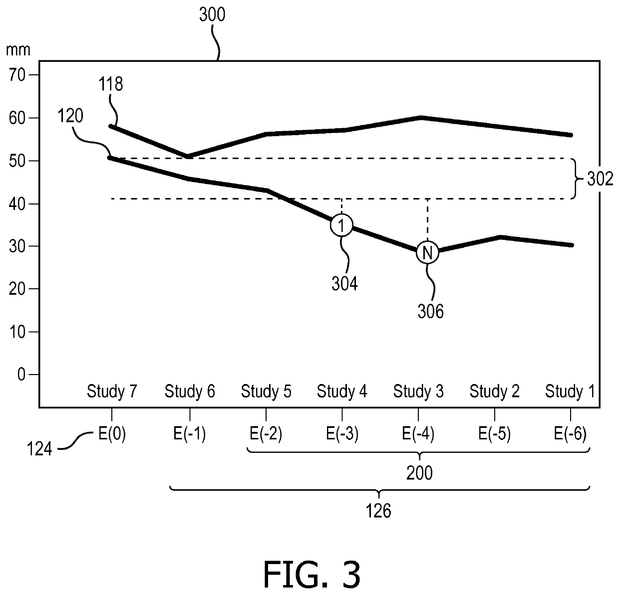
One issue with current oncological practice is the possible presence of “creep,” which is the appearance of the lesion as a “stable disease” between any two consecutive examinations, but unequivocally growing in a larger time span.

Method used


Image

  • Tumor tracking with intelligent tumor size change notice
  • Tumor tracking with intelligent tumor size change notice
  • Tumor tracking with intelligent tumor size change notice

Examples


Embodiment Construction

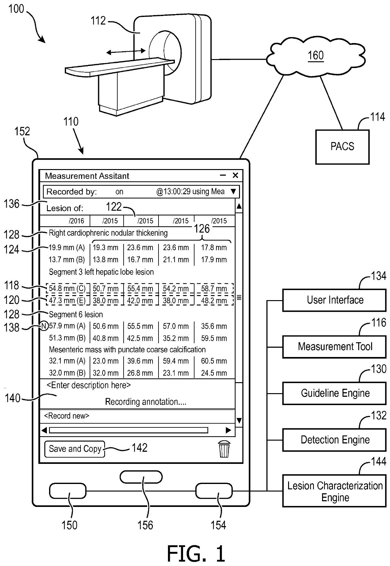
[0017]With reference to FIG. 1, an embodiment of a medical imaging system 100 with a tumor tracking device 110 is schematically illustrated. A medical image of a subject can be generated and received directly from a medical imaging scanner 112, such as a computed tomography (CT) scanner, a magnetic resonance (MR) scanner, a positron emission tomography (PET) scanner, single photon emission computed tomography (SPECT) scanner, ultrasound (US) scanner, combinations thereof, and the like. The medical image can be stored in and received from a storage subsystem 114, such as a Picture Archiving and Communication System (PACS), radiology information system (RIS), Electronic Medical Record (EMR), Hospital Information System (HIS) and the like.

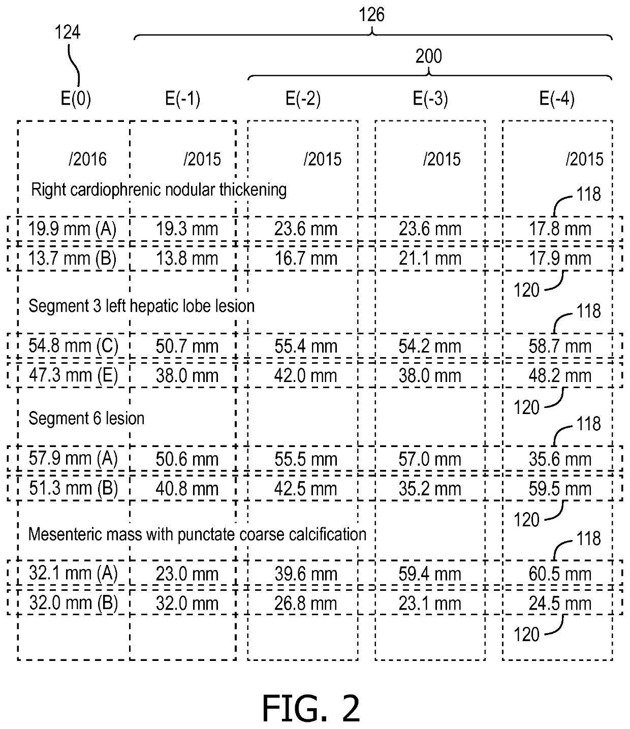
[0018]A measurement tool 116 can measure lesions in the medical image, such as a long diameter 118 of a lesion, a short diameter 120 of the lesion, and / or both. Measurements are identified chronologically. For example, measurements can include date st...


PUM


Abstract

A tumor tracking device (110) includes a guideline engine (130), a detection engine (132), and a user interface (134). The guideline engine (130) receives a current measurement and a plurality of prior measurements of at least one lesion according to a medical image of a subject, each of the current and the plurality of prior measurements identified chronologically, and calculates growth between the current measurement and a most recent measurement of the plurality of prior measurements. The detection engine (132) calculate growth between the current measurement and each of non-most recent measurements of the plurality of prior measurements, and identified at least one of the non-most recent measurements in response to calculated growth between the current measurement and each of non-most recent measurements of the plurality of prior measurements exceeding a threshold according to a medical guideline and the calculated growth between the current measurement and a most recent measurement of the plurality of prior measurements failing to exceed the threshold. The user interface (134) displays on a display device (136) an indicator (138) of the identified at least one of the non-most recent measurements of the at least one lesion.

Description

FIELD OF THE INVENTION[0001]The following generally relates to medical imaging and cancer treatment and more specifically to longitudinal tracking of lesion measurements in medical images from medical imaging modalities, such as computed tomography (CT) reconstruction, positron emission tomography (PET), single photon emission computed tomography (SPECT), magnetic resonance (MR), and combinations thereof.BACKGROUND OF THE INVENTION[0002]Interpretation of medical images can include quantitative measurements, such as the dimensions of a lesion. For example, in Response Evaluation Criteria In Solid Tumors (RECIST), a measurement of a mass lesion is taken across the lesion at a long diameter, and a measurement of a lymph node lesion is taken across the lesion at a short diameter. Both diameters are often measured, particularly for undifferentiated lesions. Guidelines, such as RECIST or those from the World Health Organization (WHO) establish standards of care in evaluating the measured ...

Claims


Application Information

Patent Timeline
Patent Type & Authority Applications(United States)
IPC IPC(8): G16H70/20G16H15/00G16H30/40G16H50/20
CPCG16H70/20G16H15/00G16H50/20G16H30/40
Inventor SEVENSTER, MERLIJNHODGE, MICHAEL STEPHEN
Owner KONINKLJIJKE PHILIPS NV
Learn More