

Selection, segmentation and analysis of exhaled breath for airway disorders assessment
a technology of exhalation and analysis, applied in the field of breath exhalation, can solve the problems of young children breathing abnormally, obstructing breathing into the mouthpiece, etc., and achieve the effect of increasing the sensitivity of the tes
- Summary
- Abstract
- Description
- Claims
- Application Information
AI Technical Summary
Benefits of technology
Problems solved by technology
Method used
Image

Examples
Embodiment Construction
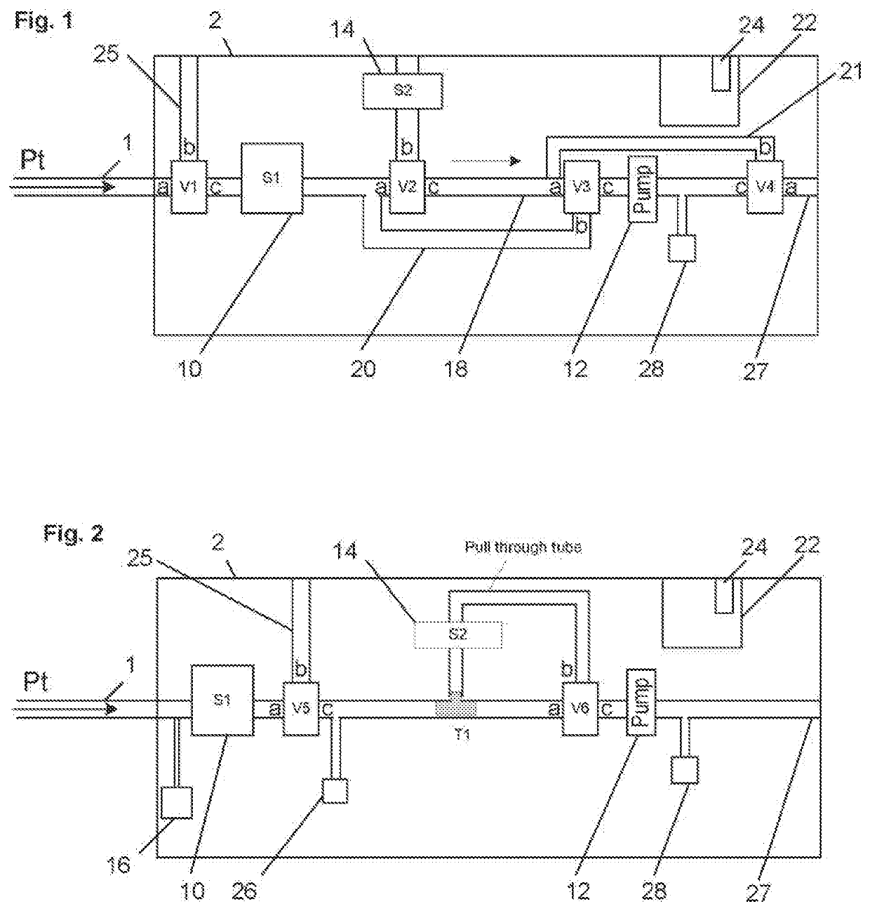
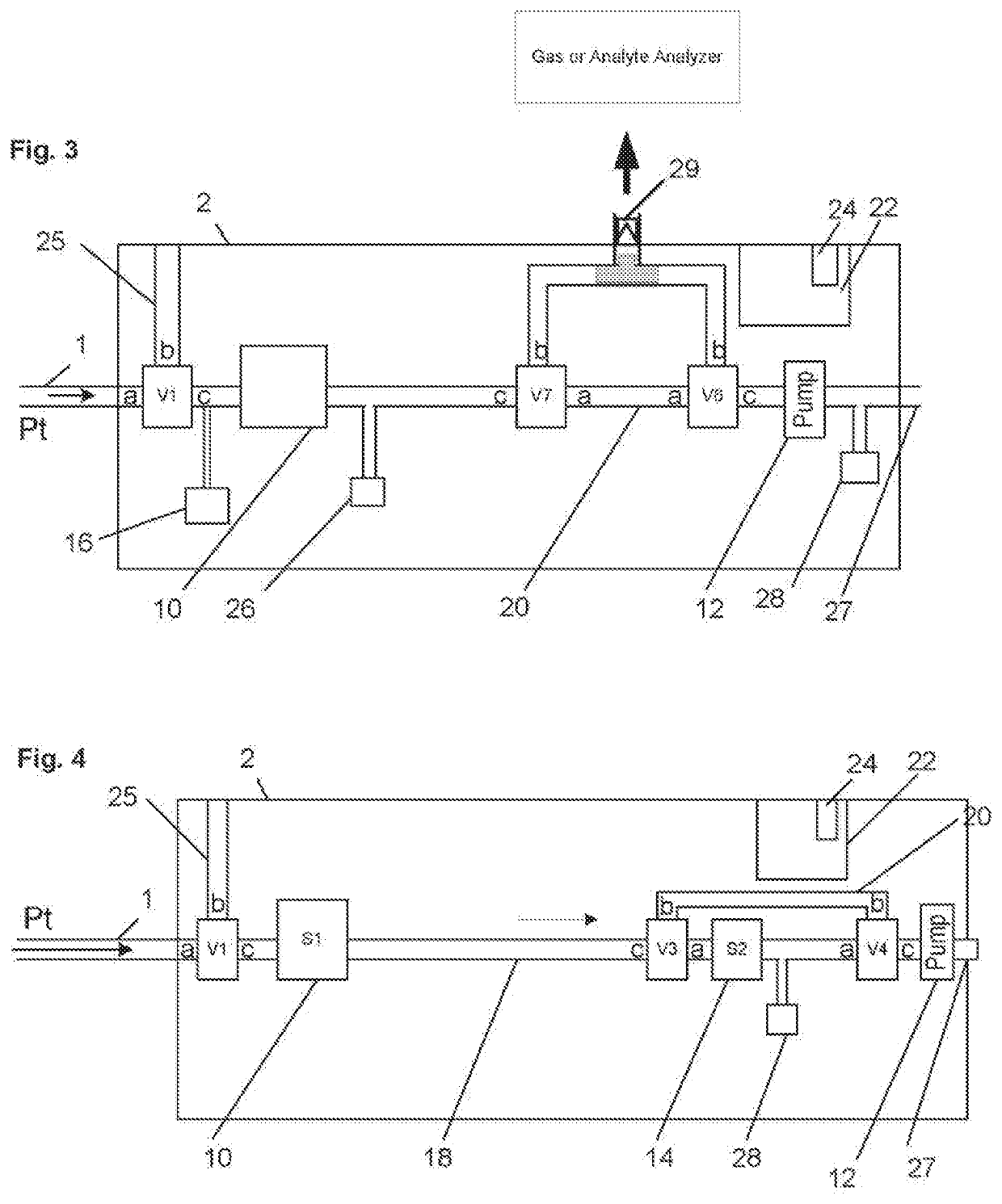
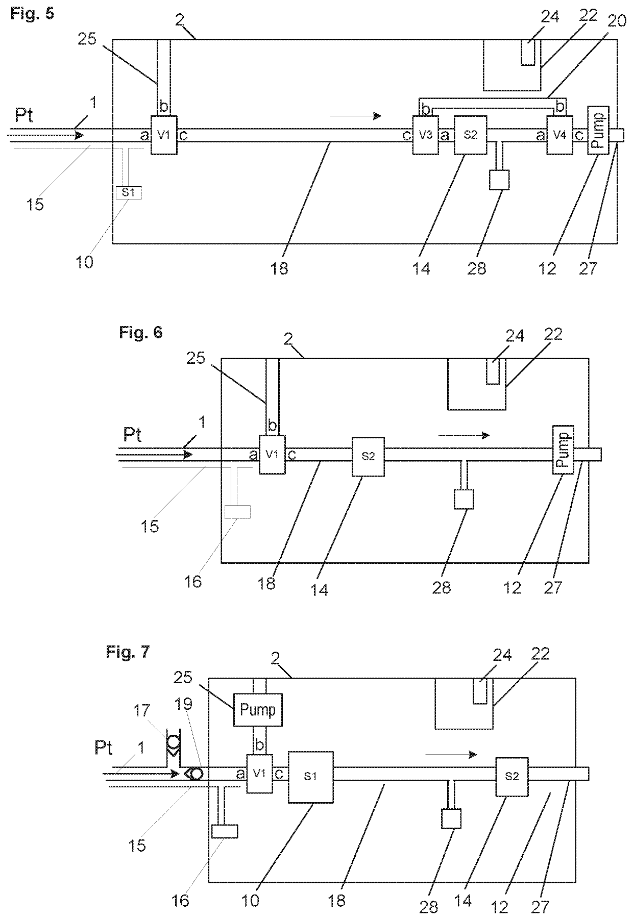
[0052]Described here are devices and methods for obtaining a gas sample for analysis especially from young children, or non-cognizant or non-compliant patients. In particular obtaining an accurate and reliable and repeatable measurement is described by automatically collecting the sample, collecting the sample in a manner that does not make the patient's breathing abnormal or invalid for the test, partitioning the expiratory gases into different partitions corresponding to different sections of the pulmonary tree in the lung, isolating a expiratory gas partition that is physiologically valid for the analysis of interest, and measuring the sample for the analyte in question. In one variation, a collection of exhaled gas is taken from a physiologically valid breath of a young child, isolating gas from the middle portion of the exhaled gas representing the gas from the middle and lower airways, and measuring that portion of gas for NO for the assessment of asthma. In the variations sho...
PUM
 Abstract
Description
Claims
Application Information