Methods of detection of compound, antibody or protein using recombinant endospores or bacteria as sensing element
a technology of recombinant endospores and sensing elements, which is applied in the field of detection methods of compound, antibody or protein using recombinant endospores or bacteria as sensing elements, can solve the problems of complex procedures, high equipment costs, and unsuitable sandwich elisa, and achieves lower detection limit/higher sensitivity, easy handling, and low cost
- Summary
- Abstract
- Description
- Claims
- Application Information
AI Technical Summary
Benefits of technology
Problems solved by technology
Method used
Image

Examples
example 1
isplay of Fusion Proteins
1-1 Plasmid Construction
[0100]The recombinant plasmids for use in the present invention were produced through conventional recombinant techniques known in the art.
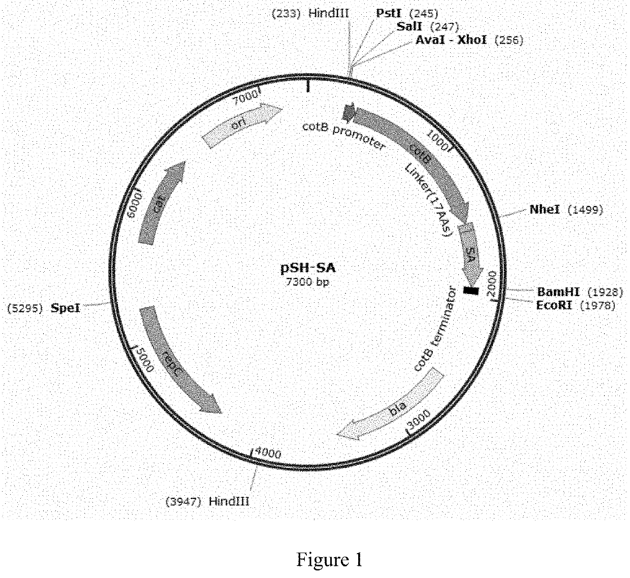
[0101]pSH-SA
[0102]CotB (Kunst et. al., 1997) and streptavidin (SA) genes were cloned into shuttle vector pMK4 (BCRC 41416, purchased from Bioresource Collection and Research Center (BCRC), Taiwan) with replication origin ori and cleavage sites for restriction enzyme HindIII, PstI, BamHI, EcoRI, NdeI and SpeI and selectable markers including Ampicillin resistance gene (AmpR(bla)) and Chloramphenicol acetyltransferase (CmR(cat)) to form recombinant plasmid pSH-SA through use of Escherichia coli DH5a (obtained from Dr. Chih-Hung Huang at National Taipei University of Technology, Taiwan) with genotype fhuA2 lacΔU169 phoA glnV44Φ80′ lacZΔM15 gyrA96 recA1 relA1 endA1thi-1 hsdR17 (Taylor et al., 1993). The cotB DNA fragment containing the promoter and structure gene without stop codon of cotB, cgtcgtcgtcg...
example 2
of Anti-Streptavidin Antibody by LFA Using Spores and Gold Nanoparticles (AuNP)
2-1 Pretreatment of Spores
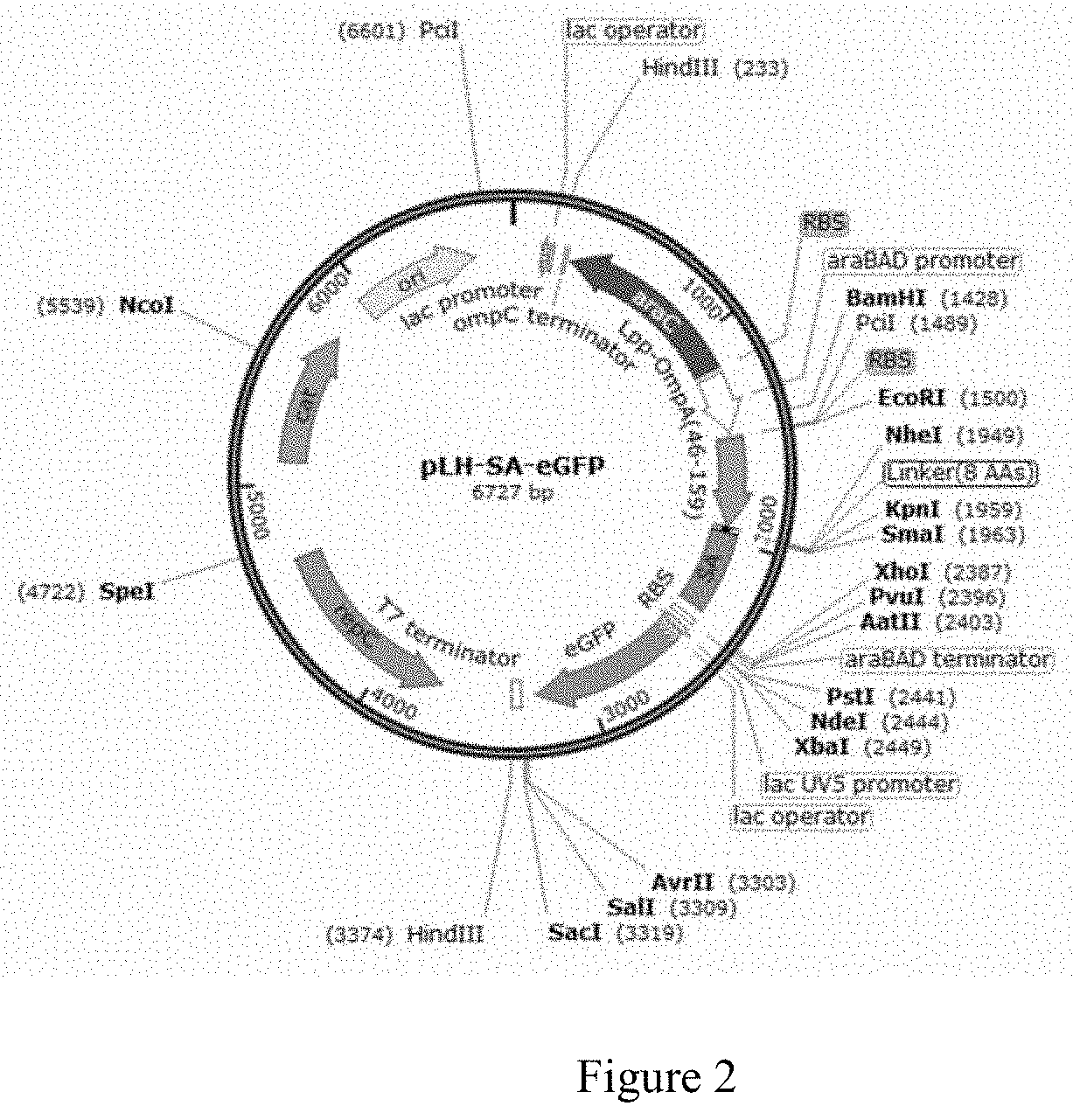
[0119]To 1000 μl of spores (B.s. 168 / pH-SA-eGFP) with OD600 of 1 was added 250 μl of 0.05% SDS and mixed for 30 minutes. The mixture was centrifuged twice (under 12000×g for 3 minutes), followed by addition of 1000 μl 1% BSA (w / v, BSA / PBS) and a mixing step for 2 hours. The spores were then centrifuged and mixed with 100 μl PBS so that the OD600 was adjusted to about 10.
2-2 AuNP Modification
[0120]100 μl of potassium carbonate solution at the concentration of 26 mM was added to 600 μl of AuNP solution (0.355 nM) (final concentration: AuNP solution at 0.3 nM; potassium carbonate solution at 3.714 mM). 1 μl of anti-spore antibody (Mybiosource / mbs612878) at the concentration of 2 mg / ml was added and the mixture was stored at 4° C. for 16 hours, followed by addition of 200 μl of 5% BSA and waiting for 30 minutes. The mixture was then centrifuged under 4000×g at 4° C. for 40 minutes. A...
example 3
of β-galactosidase by LFA Using Spores and Gold Nanoparticles (AuNP)
3-1 Pretreatment of Spores
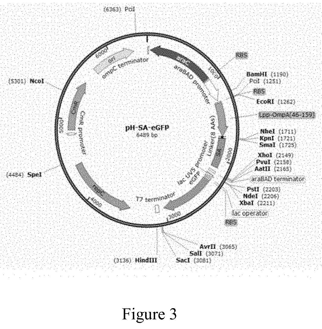
[0124]To 1000 μl of spores (B.s. 168 / pSH-SA-eGFP) with OD600 of 1 was added 250 μl of 0.05% SDS and mixed for 30 minutes. The mixture was centrifuged twice (under 12000×g for 3 minutes), followed by addition of 1000 μl 1% BSA (w / v, BSA / PBS) and a mixing step of 2 hours. The spores were then centrifuged and mixed with 100 μl PBS and the OD600 was adjusted to about 10. Afterward, 100 μl of a biotinylated anti-β-gal antibody (abcam / ab6645) at the concentration of 10−7 M was added and mixed for 2 hours, followed by centrifuging the spores and adding 100 μl of PBS so that the OD600 was adjusted to about 10.
3-2 AuNP Modification
[0125]100 μl of potassium carbonate solution at the concentration of 26 mM was added to 600 μl of AuNP solution (0.355 nM) (final concentration: AuNP solution at 0.3 nM; potassium carbonate solution at 3.714 mM). 1 μl of anti-spore antibody (Mybiosource / mbs612878) (from ra...
PUM
| Property | Measurement | Unit |
|---|---|---|
| size | aaaaa | aaaaa |
| diameter | aaaaa | aaaaa |
| volume | aaaaa | aaaaa |
Abstract
Description
Claims
Application Information
 - R&D Engineer
- R&D Manager
- IP Professional
- Industry Leading Data Capabilities
- Powerful AI technology
- Patent DNA Extraction
Browse by: Latest US Patents, China's latest patents, Technical Efficacy Thesaurus, Application Domain, Technology Topic.
© 2024 PatSnap. All rights reserved.Legal|Privacy policy|Modern Slavery Act Transparency Statement|Sitemap