Method for treating and relieving myopia
a myopia and treatment method technology, applied in the field of pharmaceutical compositions, can solve the problems of undefined mechanism of effect, high-degree myopia is a particularly dangerous visual affliction, and the economic cost of myopia has increased to us$250 million per year, so as to relieve inflammation, treat and relieve myopia, and inhibit inflammation
- Summary
- Abstract
- Description
- Claims
- Application Information
AI Technical Summary
Benefits of technology
Problems solved by technology
Method used
Image
Examples
preparation example 1
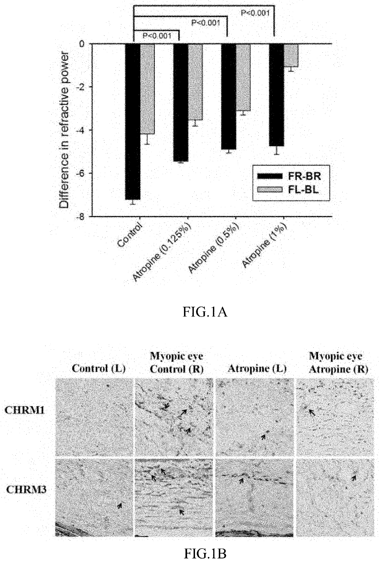
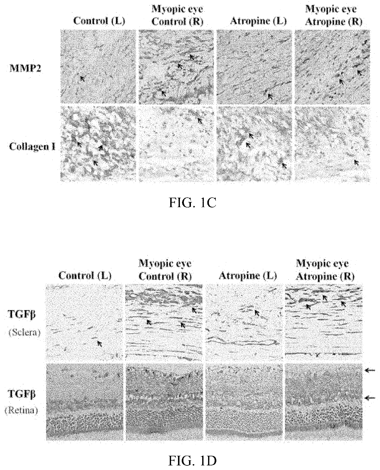
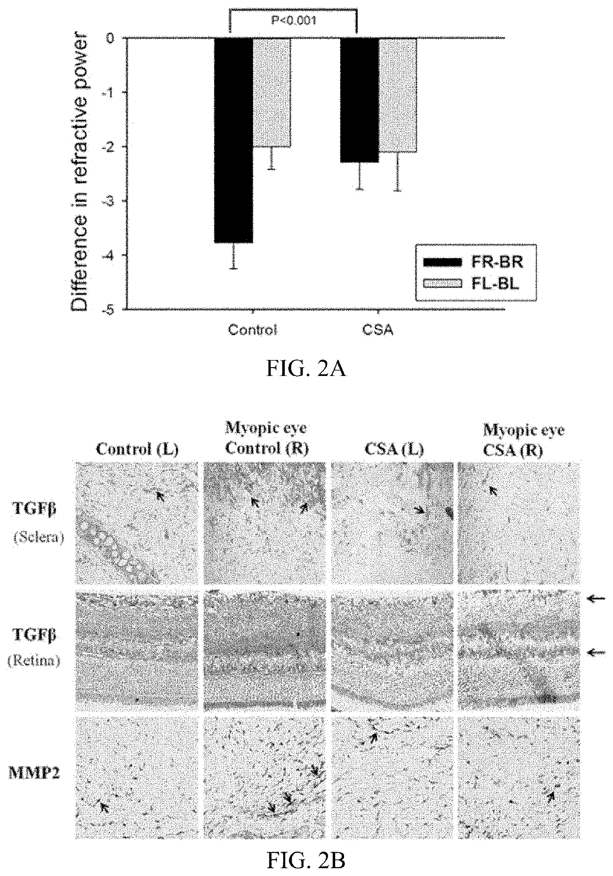
[0046]In this study, a total of 160 golden Syrian hamsters aged 3 weeks, weighing 80 to 90 g, and 20 albino guinea pigs, aged 2 to 3 weeks, were used for experiments. All animals were kept in a 12-hour light / dark cycle. All procedures were approved by the Institutional Animal Care and Use Committee of China Medical University and were conducted in accordance with the guidelines of the Use of Animals in Ophthalmic and Vision Research. Hamsters were raised with a right eyelid fusion for 21 days. Myopia was induced in the guinea pigs by covering the right eye with a cloth attached to the skin at a distance of at least 1 cm from the eye. MFD was induced in the right eye (with the left eye serving as a control) of animals randomly assigned to treatment or control groups (n=10 animals each) receiving daily applications of drug or phosphate-buffered saline (PBS), respectively, to both eyes.
preparation example 2
Cell Culture
[0047]R28 rat retinal epithelial cells were provided by Gail Seigel at the Ross Eye Institute (SUNY, Buffalo, N.Y., USA). The cells were cultured in Dulbecco's modified Eagle's medium (DMEM) with 10% fetal bovine serum (FBS) at 37° C. and 5% CO2, with medium replacement every 3 days to 4 days. Sclera were placed in a 60-mm culture dish in DMEM supplemented with 10% FBS to isolate primary scleral fibroblasts; those from fewer than three passages were used in experiments. Cells were seeded in six-well plates (1×10⁵ cells / well) and treated with lipopolysaccharide (LPS) at 100 ng / mL or left untreated for 4 hours, followed by 100 μM atropine for 24 hours. Cell lysates were collected for quantitative (q)PCR to determine gene expression levels.
preparation example 3
cDNA Microarray Analysis
[0048]Sclera tissues were obtained from eyes with or without MFD. Total RNA was isolated using the RNeasy Mini Kit (purchased from Qiagen). RNA integrity and purity were determined with an Agilent Bioanalyser. A total of five unique total RNAs were pooled together (equal amounts) for cDNA microarray analysis. cDNA microarray analysis was performed using Affymetrix GeneChip Human Genome U133 Plus 2.0 and the procedures were consistent with the manufacturer's guidelines. cDNA microarrays were scanned using a GeneArray scanner. The image files (.cel format) were analyzed using the DNA-Chip Analyser software. Genes that exhibited a differential expression greater than 1.2 fold between the control and myopic eyes were selected for ingenuity pathway analysis.
PUM
| Property | Measurement | Unit |
|---|---|---|
| volume ratio | aaaaa | aaaaa |
| volume ratio | aaaaa | aaaaa |
| weight ratio | aaaaa | aaaaa |
Abstract
Description
Claims
Application Information
 - R&D
- Intellectual Property
- Life Sciences
- Materials
- Tech Scout
- Unparalleled Data Quality
- Higher Quality Content
- 60% Fewer Hallucinations
Browse by: Latest US Patents, China's latest patents, Technical Efficacy Thesaurus, Application Domain, Technology Topic, Popular Technical Reports.
© 2025 PatSnap. All rights reserved.Legal|Privacy policy|Modern Slavery Act Transparency Statement|Sitemap|About US| Contact US: help@patsnap.com


