Method for automated unsupervised ontological investigation of structural appearances in electron micrographs

an electron micrograph and ontological investigation technology, applied in the field of automated unsupervised ontological investigation of structural appearances, can solve the problems of difficult to objectively, repeatedly and reliably identify, classify and characterize objects captured in microscopy images, and easy to miss important information in images

Active Publication Date: 2021-09-16
INTELLIGENT VIRUS IMAGING
View PDF0 Cites 0 Cited by
  • Summary
  • Abstract
  • Description
  • Claims
  • Application Information

AI Technical Summary

Problems solved by technology

When specific and predetermined detection methods are used, it is easy to miss important information in the image that are not detected by the specific detection methods.
Also, manual identification and analysis processes are difficult to carry out in a consistent way to ensure a certain quality level.
In the past, it has been difficult to objectively, repeatedly and reliably identify, classify and characterize objects captured in microscopy images.
Prior identification and classification processes of viral particles have been heavily user-biased and cumbersome to use because the prior art methods require the user to make decisions regarding how to segment images and what scales / magnification of the microscopy should be used.
Structured artificial intelligent methods that use fixed filter banks, such as state of the art convolutional neural networks, often struggle with natural alignment challenges which makes the information resolution coarser than what is optimally achievable.
Prior art solutions rely on big data sources to try to compensate for the coarseness but, as a consequence, they are often ineffective and time consuming to use for extracting new knowledge.

Method used


Image

  • Method for automated unsupervised ontological investigation of structural appearances in electron micrographs
  • Method for automated unsupervised ontological investigation of structural appearances in electron micrographs
  • Method for automated unsupervised ontological investigation of structural appearances in electron micrographs

Examples


Embodiment Construction

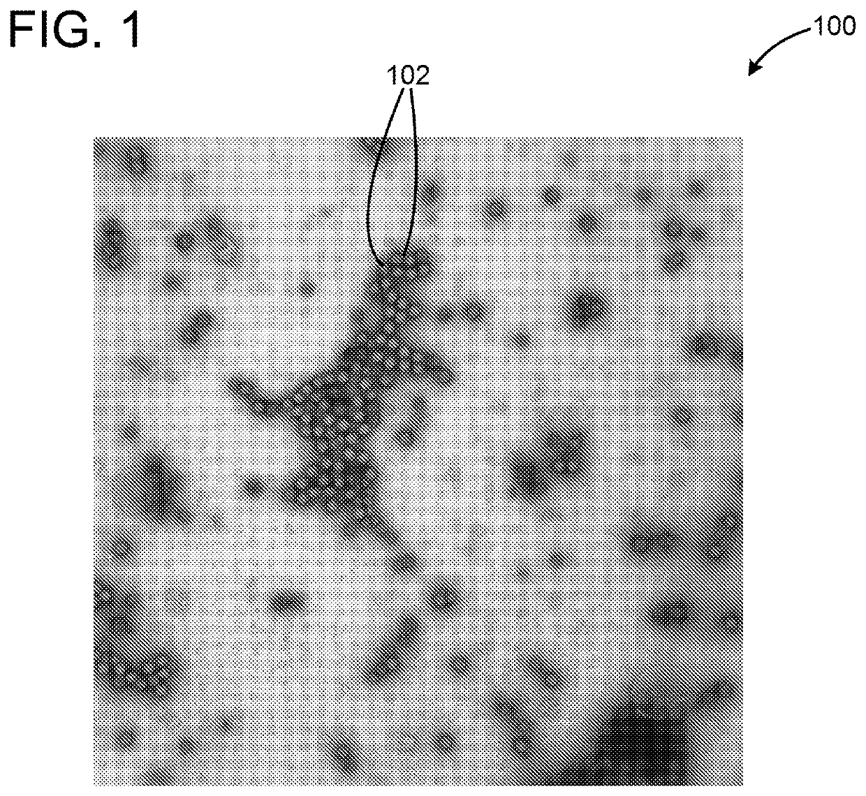
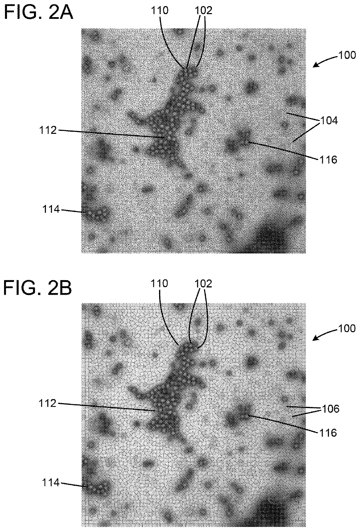
[0022]In general, the present invention is a method for (1) dividing dark data, in this case images of various types of organic or inorganic materials from electron microscopy images into meaningful segments, (2) aligning the segments so that they are meaningfully comparable, and (3) clustering the segments into classes which can be used for ontological investigation of samples that are visualized by using electron microscopy. One important aspect of the present invention is that the structural information in the image is divided into connected subsets with its own alignment fields based on the data itself and not on any fixed or predetermined structure or fixed filters which is the case for most state of the prior art methods.

[0023]In practice, a digital camera 300 (best shown in FIG. 8) is used to take a pictures or photo of an image 302 (or many images) in the electron microscope 304. The camera 300 may, of course, take many pictures of the image 302 or images. The picture is the...


PUM


Abstract

The method is for dividing dark objects, substructures and background of an image from an electron microscope into segments by analysing pixel values. The segments are transformed and aligned so that the transformed objects, sub-structures and background are meaningfully comparable. The transformed segments are clustered into classes which are used for ontological investigation of samples that are visualized by using electron microscopy. A triangle inequality comparison can be used to further cluster groups of objects to transfer understanding from different interactions between objects and to associate interactions with each other.

Description

TECHNICAL FIELD[0001]The invention relates to a method for automated unsupervised ontological investigation of structural appearances, such as organic and inorganic materials, in electron micrographs.BACKGROUND AND SUMMARY OF THE INVENTION[0002]Many efforts have been made in the past to identify, classify and analyze cell structures that include viruses and other components. Various image analysis methods have been developed to describe, segment and classify viruses by using available image technologies. Normally, the user decides beforehand what it is the user is going to search for under the microscope and at what magnification. Various detection methods have been developed in the past that are adapted to identify, measure etc. what the user is looking for. When specific and predetermined detection methods are used, it is easy to miss important information in the image that are not detected by the specific detection methods. Also, manual identification and analysis processes are d...

Claims


Application Information

Patent Timeline
Patent Type & Authority Applications(United States)
IPC IPC(8): G06K9/00G06K9/46G06K9/62
CPCG06K9/0014G06K9/6202G06K9/4661H01J2237/221G06V20/695G02B21/361G06V20/69G06V10/60G06V10/751
Inventor RYNER, MARTIN
Owner INTELLIGENT VIRUS IMAGING
Why Eureka
Try Eureka