miRNAS FOR REDUCING VENTRICLE ENLARGEMENT
a ventricle enlargement and enlargement technology, applied in the direction of nervous disorders, biochemistry apparatus and processes, drug compositions, etc., can solve the problems of difficult diagnosis of neurodevelopmental diseases such as schizophrenia (scz) and difficult reproduction of polygenic diseases in animal models
- Summary
- Abstract
- Description
- Claims
- Application Information
AI Technical Summary
Benefits of technology
Problems solved by technology
Method used
Image
Examples
example 1
and Methods
[0193]Animals. Both male and female mice (2-9 months old) were used for all experiments. The generation of Df(16)1 / +, Dgcr8+ / −, Arl13beGFP, Foxj1Cre, and Ai14 mouse lines has been reported previously (Delling et al., 2013; Earls et al., 2012; Lindsay et al., 1999; Madisen et al., 2010; Zhang et al., 2007). Df(16)1 / + and Dgcr8+ / − mouse strains were back-crossed onto the C57BL / 6J genetic background for at least 10 generations. The care and use of animals were reviewed and approved by the St. Jude Children's Research Hospital (St. Jude) Institutional Animal Care and Use Committee.
[0194]Magnetic Resonance Imaging. The animal MRI study was performed using a 7 T Bruker ClinScan system (Bruker BioSpin MRI GmbH, Germany) equipped with a 12S gradient coil. A mouse head volume coil (Bruker BioSpin) was used. Animals were anesthetized and maintained with 1.5%-2% isoflurane during the experiments. Transverse T2-weighted turbo spin echo images were acquired for volume measurements (Re...
example 2
fficiency of the 22q11DS Gene Dgcr8 Causes Progressive Ventriculomegaly
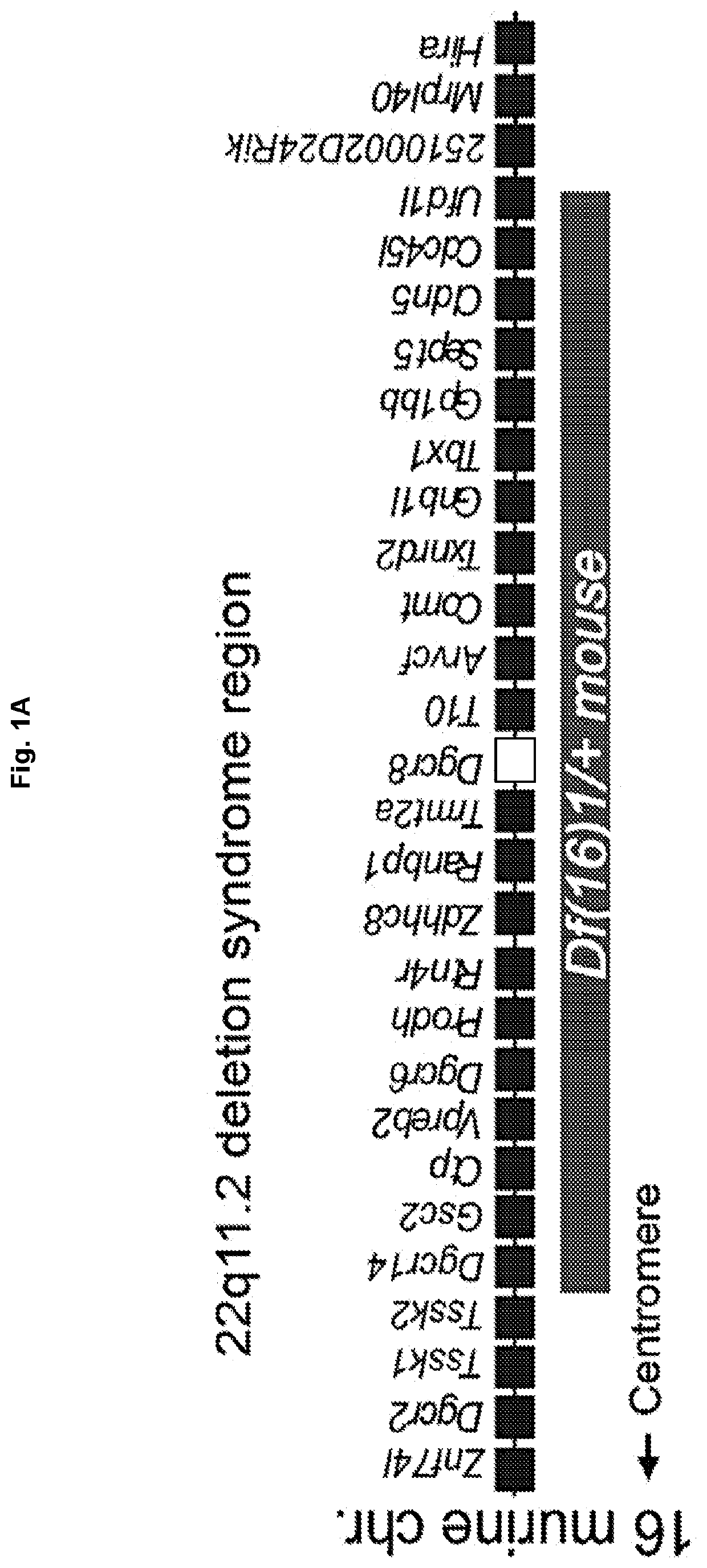
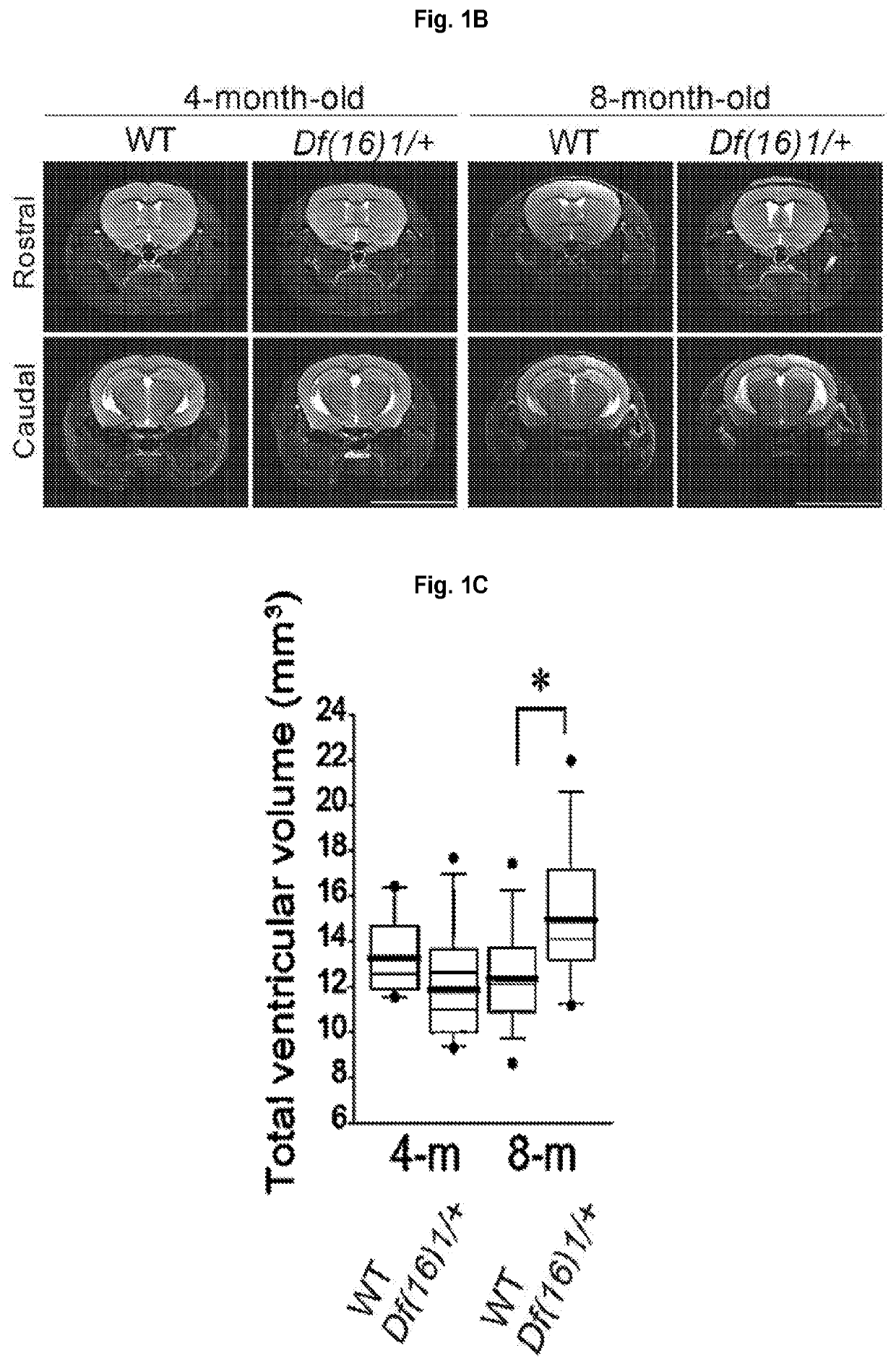
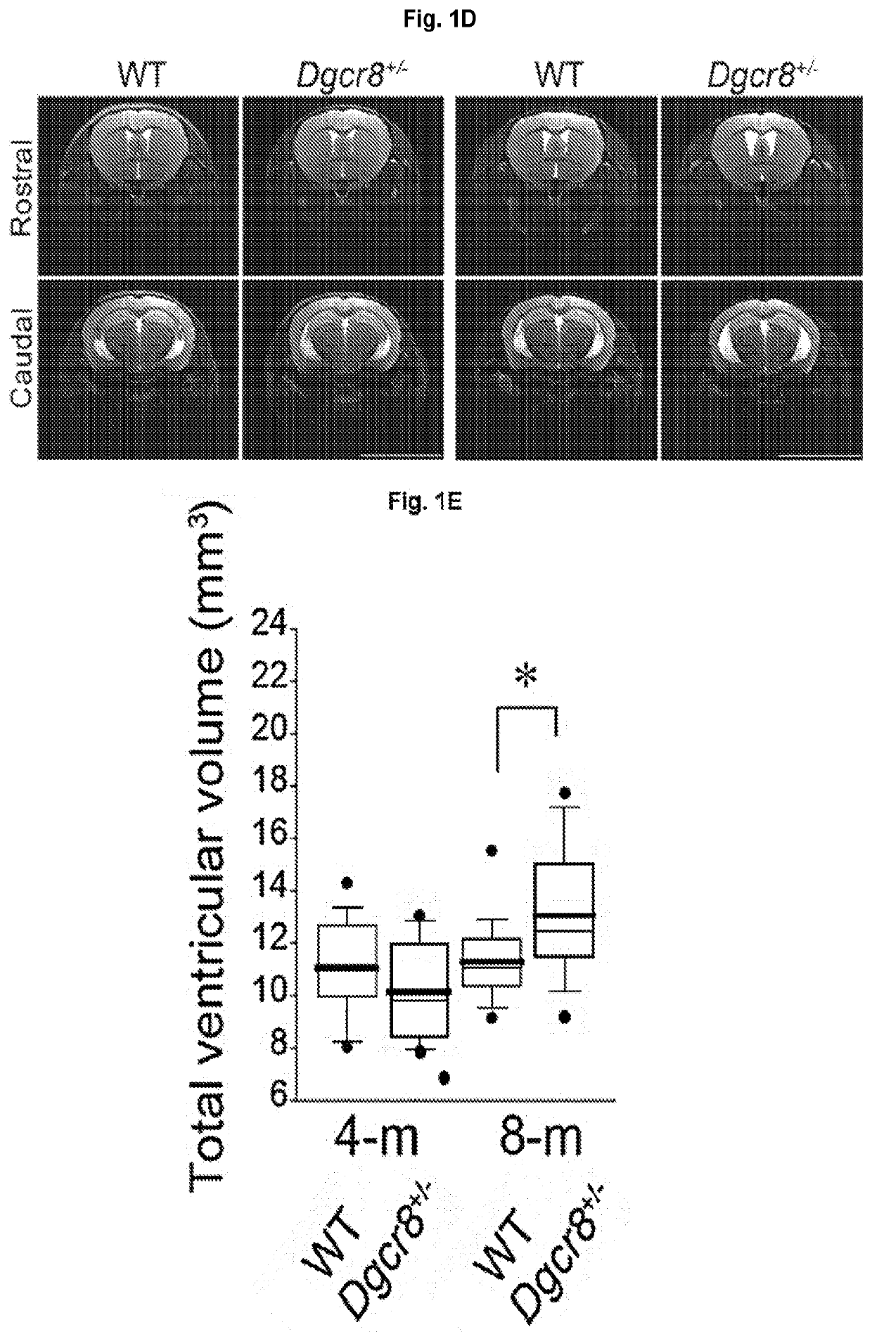
[0215]Magnetic resonance imaging (MRI) analysis was performed in Df(16)1 / + murine models of 22q11DS (Lindsay et al., 1999) (FIG. 1A). The onset of clinical manifestations of psychosis in patients with 22q11DS or SCZ typically occurs during late adolescence or early adulthood [16-30 years (Almeida et al., 1995; Bassett et al., 2003; Mueser and McGurk, 2004)]. This age is developmentally equivalent to 3 to 4 months in mice (Flurkey et al., 2007). Thus, the neuroanatomical features were examined in mice that were 4 months or older. A considerable increase in the total ventricular volume was detected in 8-month-old but not 4-month-old Df(16)1 / + mice compared to wild-type (WT) littermates (FIGS. 1B, 1C). At the regional level, a significant enlargement of the LVs and TV was observed but there was no difference in the size of the fourth ventricle or the aqueduct in 8-month-old Df(16)1 / + mice compared to age matched con...
example 4
loinsufficiency does not Affect Ependymal Cell Structure or Planar Polarity
[0221]The increase in ventricular volume in Dgcr8+ / − mice was not associated with structural changes in ependymal cilia or planar cell polarity. Transmission electron microscopy (TEM) revealed normal microtubule configuration in the 9+2 axoneme and basal body docking to the apical membrane of ependymal cells in Dgcr8+ / − mice (FIGS. 3A-3D). Scanning electron microscopy also showed no abnormalities in morphology, length, or the number of motile cilia on the wall of the LVs in the Dgcr8+ / − mice compared to WT littermates (FIGS. 3E-3H, 3M, 3N). Furthermore, planar polarity of ependymal cells was not altered in Dgcr8+ / − mice (FIGS. 3I-3L). Planar polarity within the ependymal cells is determined by the location of the microtubule-based basal bodies, which give rise to the motile cilia (Mirzadeh et al., 2010a). Co-labeling of the cell adherens junction and basal body with antibodies against β-catenin and γ-tubulin ...
PUM
| Property | Measurement | Unit |
|---|---|---|
| angles | aaaaa | aaaaa |
| number-average molecular weight | aaaaa | aaaaa |
| number-average molecular weight | aaaaa | aaaaa |
Abstract
Description
Claims
Application Information
 - R&D
- Intellectual Property
- Life Sciences
- Materials
- Tech Scout
- Unparalleled Data Quality
- Higher Quality Content
- 60% Fewer Hallucinations
Browse by: Latest US Patents, China's latest patents, Technical Efficacy Thesaurus, Application Domain, Technology Topic, Popular Technical Reports.
© 2025 PatSnap. All rights reserved.Legal|Privacy policy|Modern Slavery Act Transparency Statement|Sitemap|About US| Contact US: help@patsnap.com


