Surgical apparatus and method

a technology of surgical equipment and wall tissue, applied in the field of surgical equipment and methods, can solve the problems of pathologist possibly providing incorrect or incomplete studies, unable to resect mural disease, limited endoscopic polypectomy, etc., to reduce the chance of contact, facilitate the complete removal of wall tissue, and reduce the effect of effectively minimizing the opportunity

Inactive Publication Date: 2001-07-24
MCGUCKIN JR JAMES F
View PDF45 Cites 2565 Cited by
  • Summary
  • Abstract
  • Description
  • Claims
  • Application Information

AI Technical Summary

Benefits of technology

The technical effect of this patent is that it describes an improved method and device for removing abnormal tissue growths such as polyps or cancerous tumors from bodily cavities like colonoscopies. One key feature is that the device can remove all layers of tissue without leaving any behind, which reduces the risk of disease recurrence. Additionally, the device design allows for easy cleaning and disinfecting between uses.

Problems solved by technology

The technical problem addressed in this patent is how to improve the ability of surgeon's tools to resect and anastomozse diseased tissue without causing damage or compromising their effectiveness while maintaining minimal invasivity. Current methods require large laparotomy incisions or expensive and potentially unsafe laparoscopic procedures, limiting the benefits of endoscopy. There is a need for new technology to address these issues.

Method used


Image

  • Surgical apparatus and method
  • Surgical apparatus and method
  • Surgical apparatus and method

Examples


Embodiment Construction

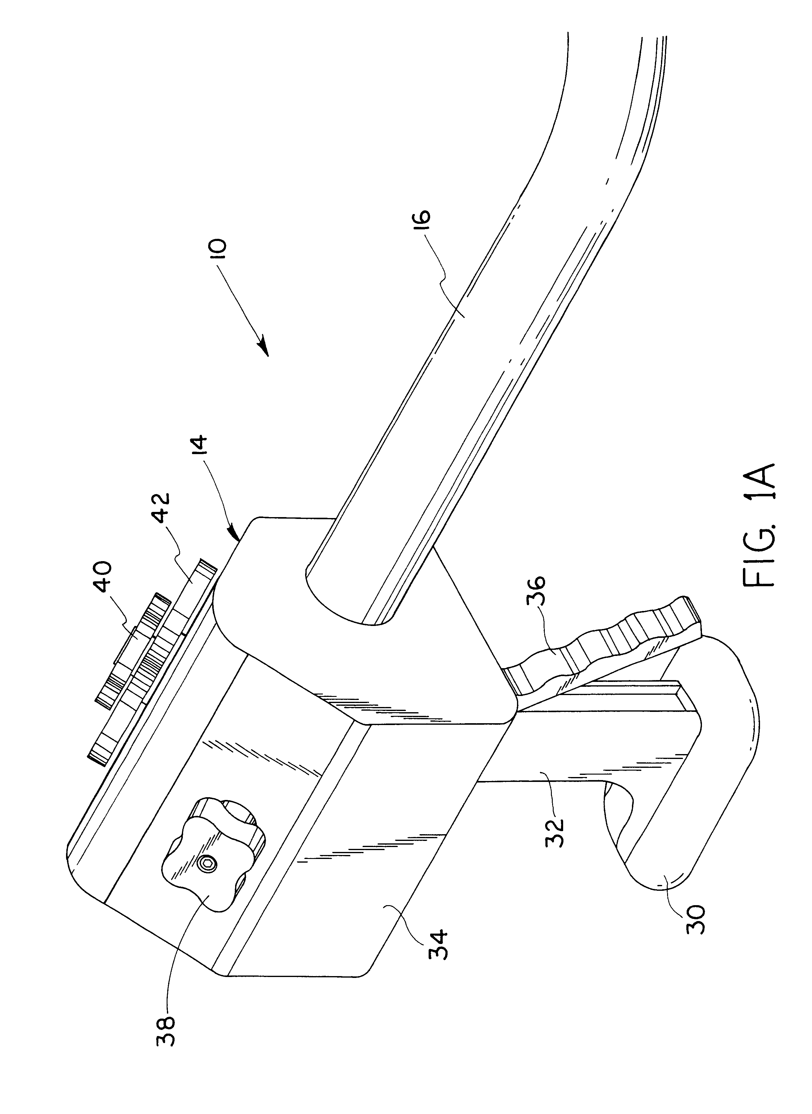
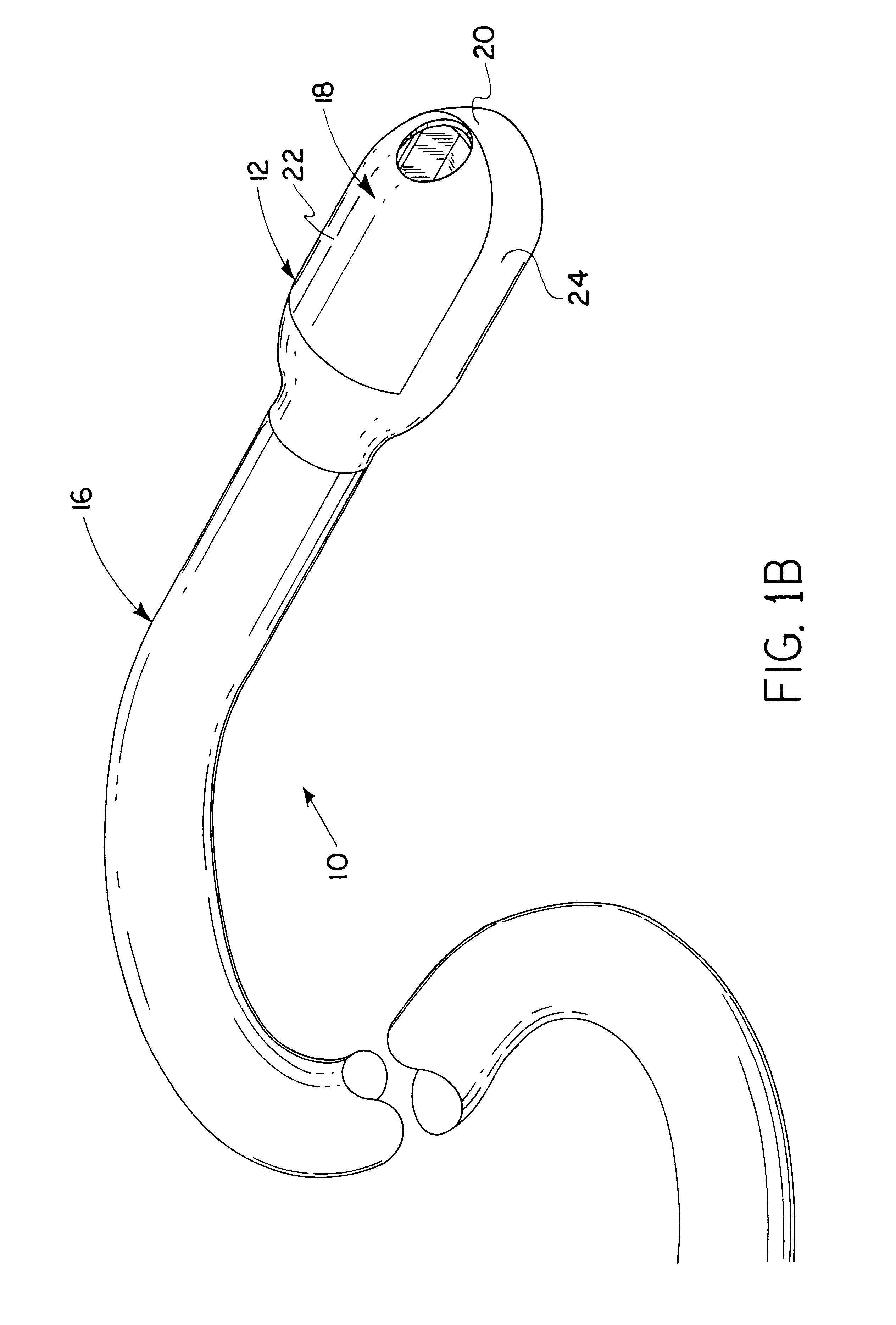
Referring to the drawings in general and to FIGS. 1A and 1B in particular, apparatus for removing malignant, other diseased or otherwise undesirable tissue from a lumen wall, such as the colon wall, while within a lumen, such as the colon, is designated generally 10 and includes a longitudinally elongated operating capsule designated generally 12, an operating control module designated generally 14 and a cable carrying flexible tubular member, designated generally 16, connecting operating capsule 12 and operator control module 14. Operating capsule 12 preferably includes an outer shell 18 having a preferably curved end 20 at one longitudinal extremity thereof which is also a longitudinal extremity of apparatus 10.

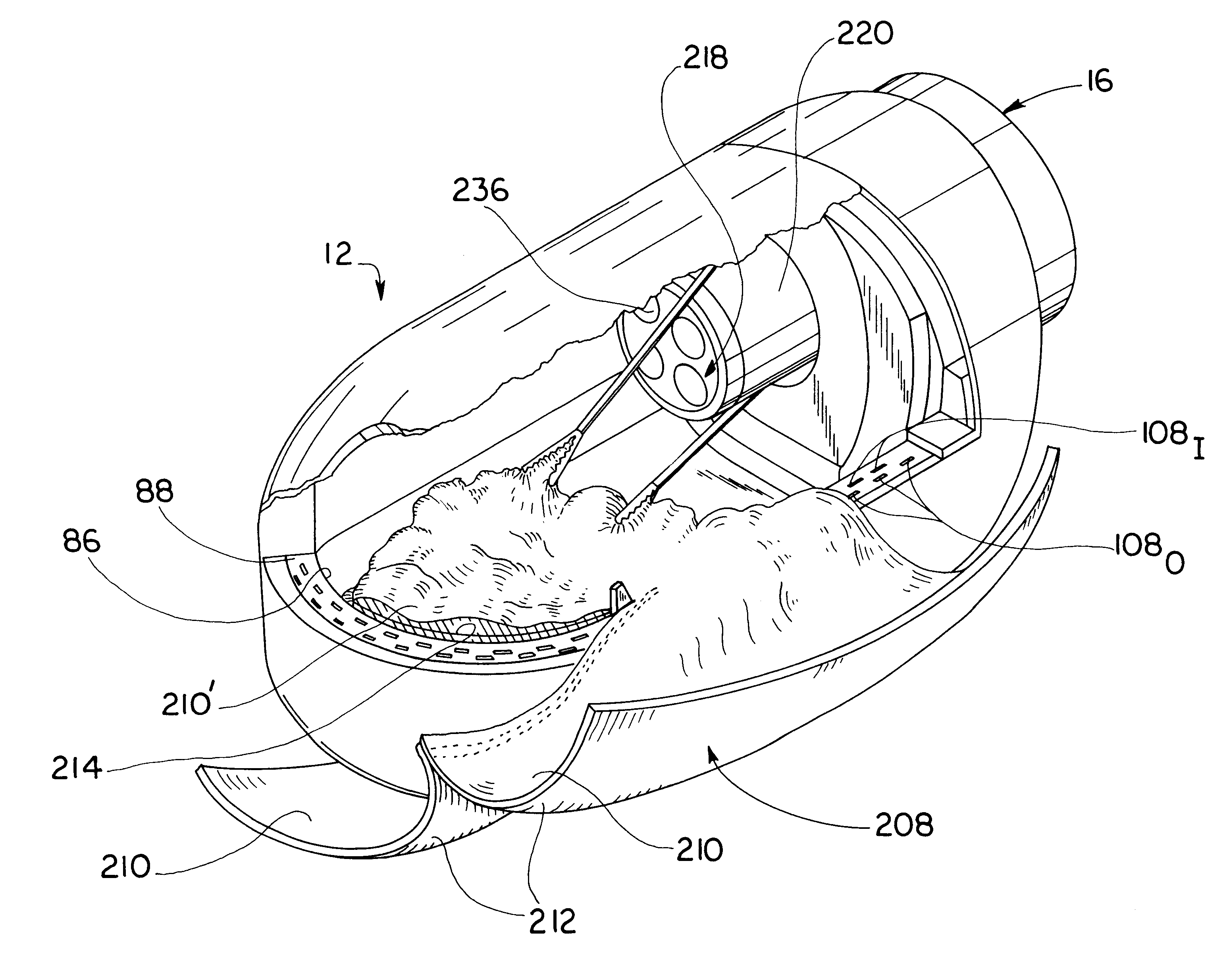
FIG. 2 illustrates the manner in which a preferred embodiment of the apparatus from removing malignant, other diseased or otherwise undesirable tissue from a lumen wall, which apparatus is designated generally 10 and is illustrated in FIGS. 1A and 1B, is preferably used in ...


PUM

PropertyMeasurementUnit
Angleaaaaaaaaaa
Lengthaaaaaaaaaa
Forceaaaaaaaaaa

Abstract

Surgical method and apparatus for resectioning tissue, preferably lumenal tissue, with a remaining portion of an organ being anastomized with staples or other fastening device, preferably endolumenally. The apparatus may be inserted via a naturally occurring body orifice or a surgical incision and then advanced using either endoscopic or radiological imaging guidance to an area where surgery is to be performed. Under endoscopic or diagnostic imaging guidance the apparatus is positioned so tissue to be resected is manipulated into an inner cavity of the apparatus. The apparatus then cuts the diseased tissue after stapling and retains the diseased tissue within the apparatus. The rent resulting in a border of healthy tissue is anastomosed with surgical staples.

Description


Claims


Application Information

Patent Timeline
Owner MCGUCKIN JR JAMES F
Why Eureka
Try Eureka