Urethra surgical device

a surgical device and urethra technology, applied in the field of medical devices, can solve problems such as loss of bladder control

Inactive Publication Date: 2002-08-13
ARTES MEDICAL USA
View PDF6 Cites 89 Cited by
  • Summary
  • Abstract
  • Description
  • Claims
  • Application Information

AI Technical Summary

Benefits of technology

The urethra scope of the present invention provides lighting and a clear path to all urethral tissues and the bladder sphincter. The filler material may comprise collagen and / or micro-spheres, such as disclosed in U.S. Pat. No. 5,344,452, or, for example, any other type of injectable bulking agent. In accordance with the present invention, the injection of a urinary bulking agent into the respective tissues serves to fortify the respective tissue structures and re-establish bladder control. The urethra scope and associated methods of operation disclosed herein may be configured for and used on other body passages as well in modified embodiments. The entire injection procedure is preferably performed on an outpatient basis requiring minimal or no sedation of the patient.
The urethra scope is designed to be a tissue expansion and visualization device for insertion into tissues and tissue lumens, such as the female urethra. The urethra scope can be manufactured as a reusable or disposable medical instrument or a combination of both. In a preferred embodiment, the device is constructed with a disposable insertion probe (single-use, sterile item comprising or consisting of, for example, plastic, stainless steel, or wire coated with, for example, a polymer or vitreous material), which is attached to a reusable handle (limited use item). The wire construction may comprise, for example, a mesh-shaped conical configuration, for example, wherein the wire is coated with a biocompatible material. In addition to serving as a tissue expansion and visualization tool, the urethra scope can serve as a diagnostic tool to enable physicians to treat their patients with injection treatments, surgical ablation of tissue, tissue biopsies, etc. The advantages of the urethra scope include cost effectiveness, ease of use, decreased likelihood of cross contamination due to sterile disposable cones that are inserted into the urethra, and improved ergonomics and handling. The placement of tangential and distal windows in the insertion probe allows for visualization and treatment of the entire length and circumference of the urethral walls and bladder neck.

Problems solved by technology

In a patient having this condition, tissue relaxation of the sphincter mechanism, located at the urinary outflow of the bladder into the urethra, can cause a loss of bladder control.

Method used


Image

  • Urethra surgical device
  • Urethra surgical device
  • Urethra surgical device

Examples


Embodiment Construction

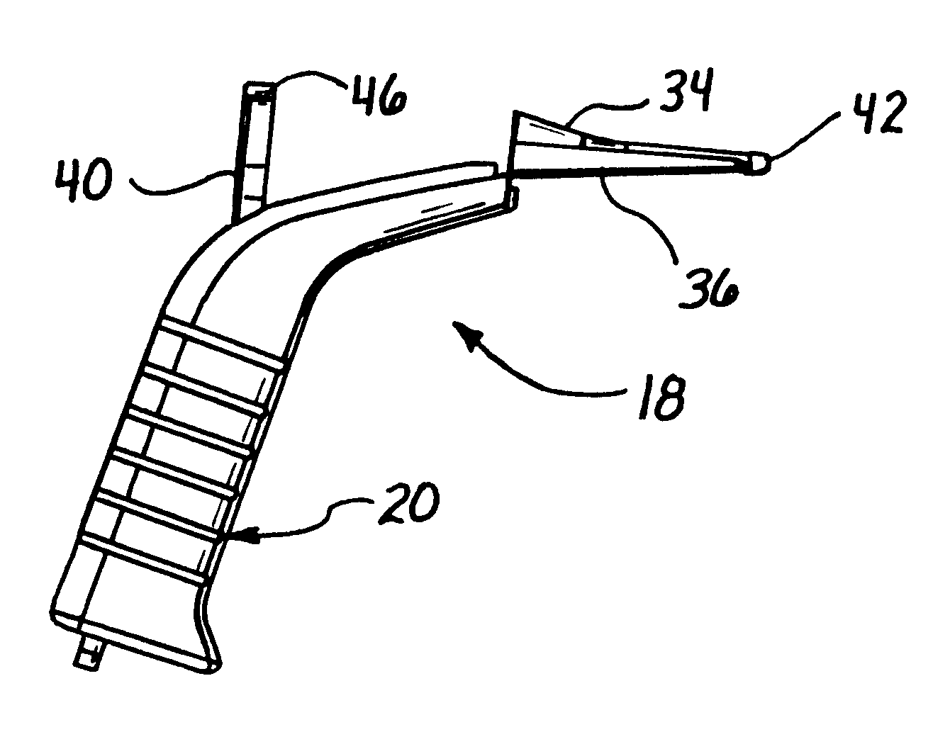
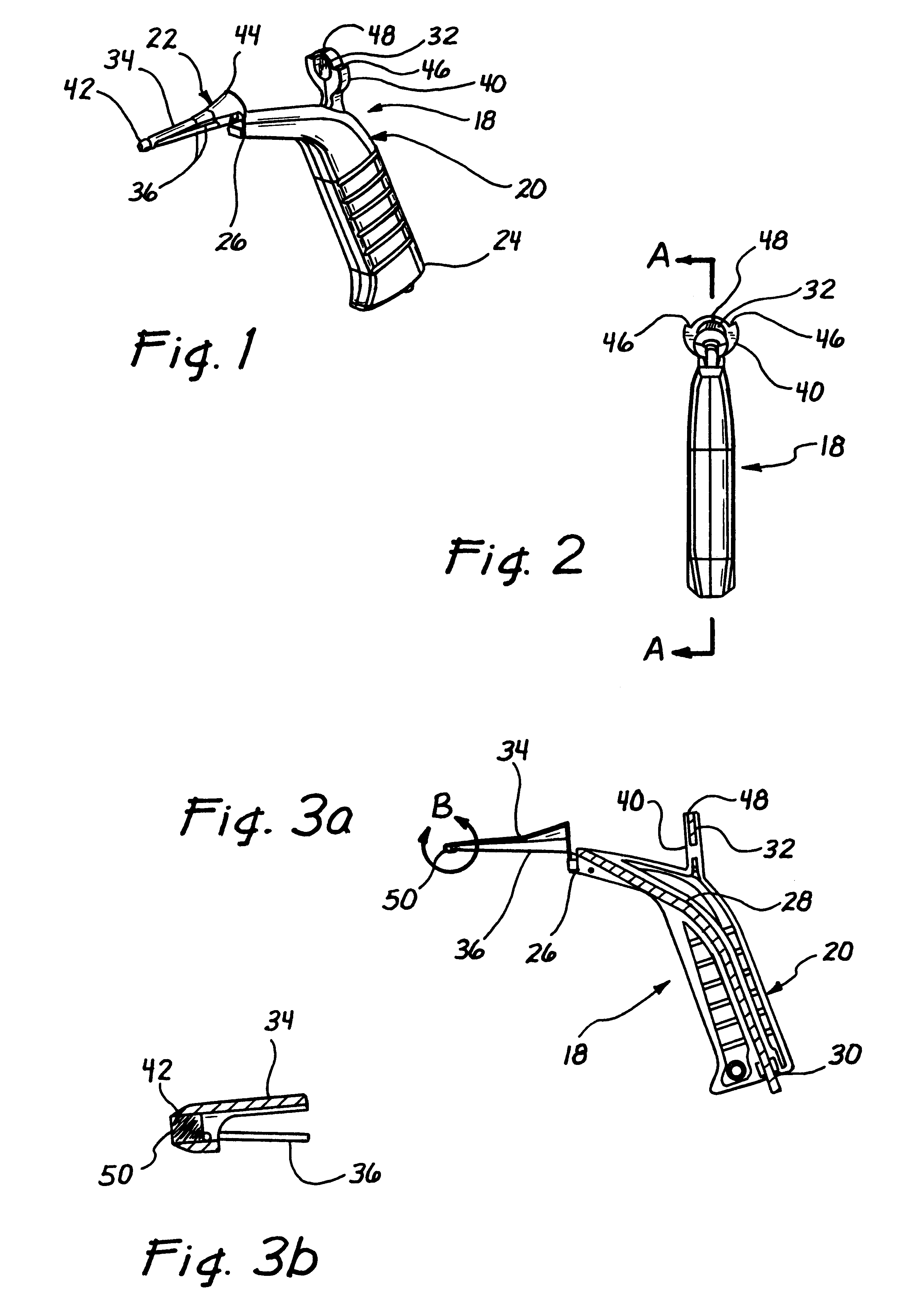
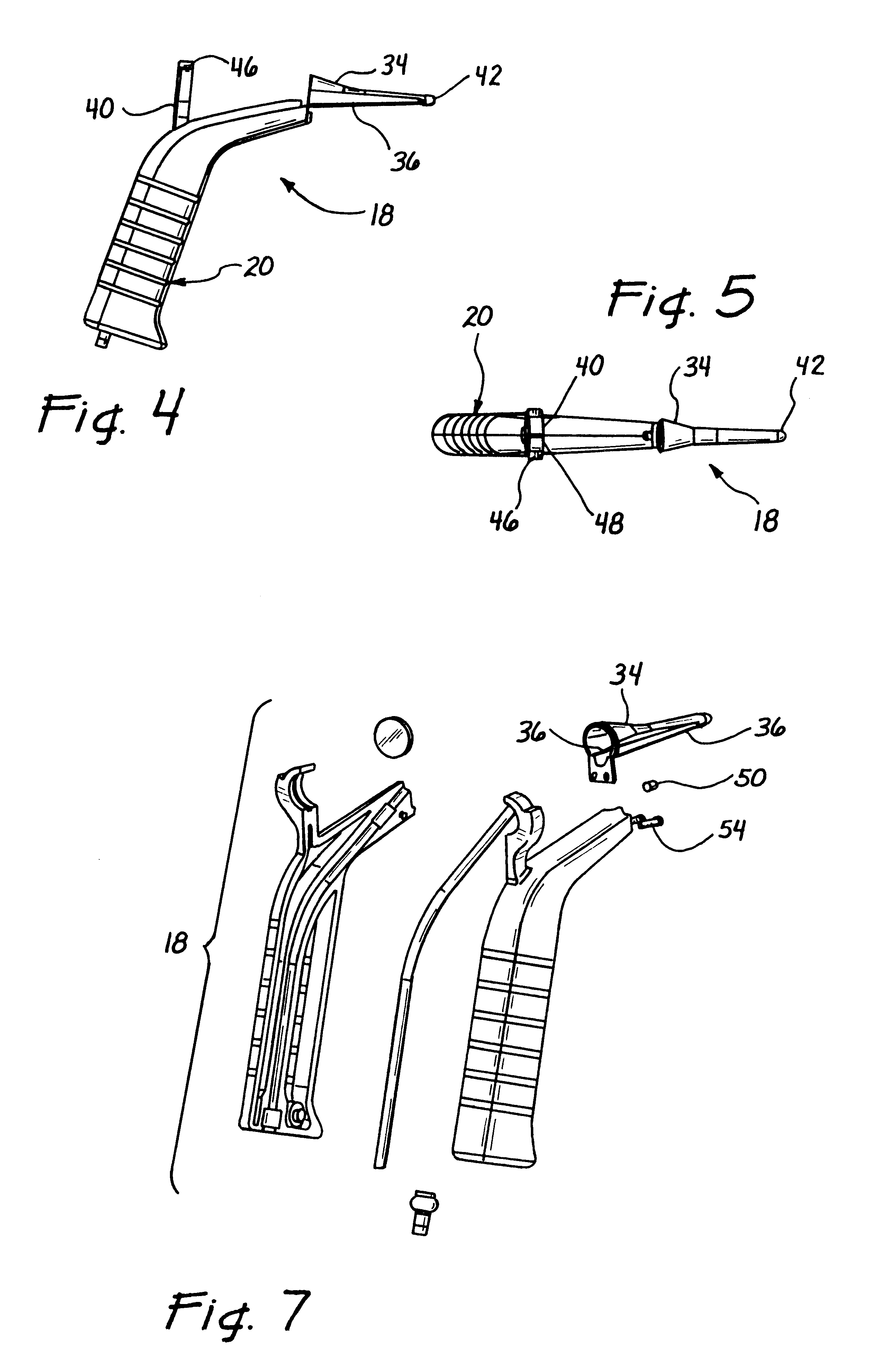
Referring more particularly to the drawings, FIG. 1 illustrates a urethra scope 18 in accordance with the present invention comprising a handle 20 and an insertion probe 22. The handle 20 comprises a proximal end 24 and a distal end 26, and the insertion probe 22 is removably connected to the distal end 26 of the handle 20. FIG. 2 illustrates a front elevation view of the urethra scope 18. The handle 20 of the urethra scope preferably comprises molded plastic and in the illustrated embodiment comprises a "pistol grip" style but is not limited to this construction. Other grip shapes, such as a simple round handles similar to the handles of standard laryngoscopes and otoscopes, may be used as well in modified embodiments. As an alternative to molded plastic, the handle 20 can be machined or formed out of surgical stainless steel for increased durability.

A lens 32 is connected to the handle 20 for providing a magnified view into the insertion probe 22 to aid the vision of a user during...


PUM


Abstract

A surgical device for expanding and viewing tissue is disclosed. The surgical device includes a handle, an insertion probe, and at least one window disposed within the insertion probe. The handle has a proximal end and a distal end, and the handle is sized and shaped to be held by a surgeon. The insertion probe has a proximal end, a distal end, and a longitudinal axis extending between the proximal end and the distal end. The proximal end of the insertion probe is removably attached to the distal end of the handle. The window extends along a length of the longitudinal axis, and has an opening that is sufficient in size to allow tissue to prolapse through the window and into the insertion probe.

Description

1. Field of the InventionThe present invention relates generally to medical devices and, more particularly, to surgical tools and scopes for viewing and performing surgical operations within body passages.2. Description of Related ArtA condition known as stress urinary incontinence refers to a functionally insufficient urinary tract of a patient. In a patient having this condition, tissue relaxation of the sphincter mechanism, located at the urinary outflow of the bladder into the urethra, can cause a loss of bladder control. A filler material, such as collagen, can be injected into and adjacent to the urinary sphincter muscle at the bladder neck to "bulk" up the muscle tissue and help ensure adequate closure of the urinary sphincter.In accordance with the present invention, a hand-held device or "urethra scope" is provided that is used to expand and visualize tissue within a body lumen. When the body lumen comprises a female urethra, the urethra scope is inserted through the urethr...

Claims


Application Information

Patent Timeline
Patent Type & Authority Patents(United States)
IPC IPC(8): A61B1/307A61B1/32A61B17/34A61B19/00A61B17/02A61B17/00G02B23/24A61B1/303A61B1/31A61M5/14
CPCA61B1/00105A61B1/06A61B1/307A61B1/32A61B17/0206A61B2017/00296A61B2017/0046A61B2017/3405A61B90/361
Inventor LEMPERLE, STEFAN M.STONE, CORBETTKELLEHER, BRIANLIPSITZ, DAVID
Owner ARTES MEDICAL USA
Why Eureka
Try Eureka