Density nodule detection in 3-D digital images

a density nodule and digital image technology, applied in the field of computer-aided detection of abnormal lesions and features in digital images, can solve problems such as speed and accuracy in processing, and achieve the effect of fast and effective overall algorithm and quick identification of regions

Inactive Publication Date: 2005-06-21
MEVIS MEDICAL SOLUTIONS
View PDF8 Cites 108 Cited by
  • Summary
  • Abstract
  • Description
  • Claims
  • Application Information

AI Technical Summary

Benefits of technology

[0020]A first stage uses digital image processing techniques to quickly highlight regions requiring further and more detailed processing. The first stage operates with somewhat lower sensitivity than is possible with other more detailed analyses. In this way, computation resources can be more effectively applied to areas of concern.
[0022]Since the first stage effectively and quickly identifies regions for further processing, the combination of the first stage analysis with the additional processing of second stage results in a fast yet effective overall algorithm to identify potential anatomical abnormalities from digital volumes.

Problems solved by technology

Key issues in digital image processing are speed and accuracy.

Method used


Image

  • Density nodule detection in 3-D digital images
  • Density nodule detection in 3-D digital images
  • Density nodule detection in 3-D digital images

Examples


Embodiment Construction

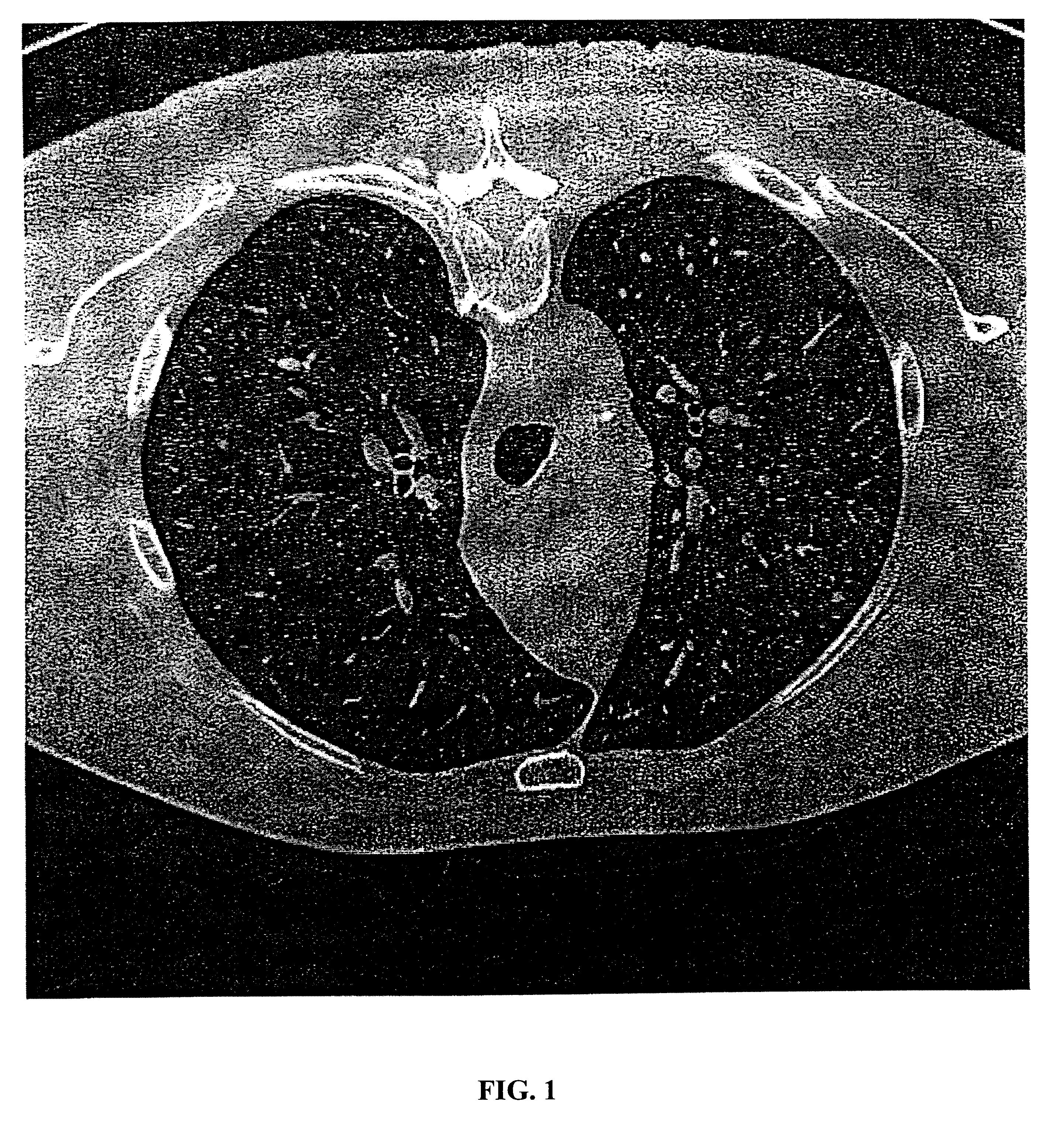
[0034]The present invention provides for systems and methods capable of effective and accurate detection of suspicious features identified from 2-D and 3-D digital images. A 3-D digital volume can be generated from the 2-D slices by any one of various techniques known in the art. The terms “digital” and “digitized” as used herein will refer to images or volumes, as appropriate, in a digital or digitized format acquired via a digital acquisition system or via conversion from an analog image.

[0035]The digital image sections and volumes to be processed, rendered, displayed or otherwise used include digitized images acquired through any plane, including, without limitation, saggital, coronal and axial (or horizontal, transverse) planes and including planes at various angles to the saggital, coronal or axial planes. While the disclosure may refer to a particular plane or section, such as an axial section or plane, it is to be understood that any reference to a particular plane is not nec...


PUM


Abstract

An algorithm is quickly scans a digital image volume to detect density nodules. A first stage is based on a transform to quickly highlight regions requiring further processing. The first stage operates with somewhat lower sensitivity than is possible with more detailed analyses, but operates to highlight regions for further analysis and processing. The transform dynamically adapts to various nodule sizes through the use of radial zones. A second stage uses a detailed gradient distribution analysis that only operates on voxels that pass a threshold of the first stage.

Description

[0001]This application is a continuation-in-part of co-pending application Ser. No. 09 / 946,209 filed Sep. 5, 2001 which is a CIP of application Ser. No. 09 / 907,585 filed Jul. 17, 2001 which in turn is a continuation of Ser. No. 09 / 103,290 filed on Jun. 23, 1998, now U.S. Pat. No. 6,263,092, which in turn is a continuation of Ser. No. 08 / 676,660, filed on Jul. 10, 1996, now U.S. Pat. No. 5,815,591.CROSS-REFERENCE TO RELATED APPLICATIONS[0002]Related applications are:[0003]“Method and System for the Display of Regions of Interest in Medical Images,” Ser. No. 09 / 990,508, filed Nov. 21, 2001;[0004]“Vessel Segmentation with Nodule Detection,” filed concurrently herewith;[0005]“Automated Registration of 3-D Medical Scans of Similar Anatomical Structures,” filed concurrently herewith;[0006]“Lung Field Segmentation From CT Thoracic Images,” filed concurrently herewith;[0007]“Pleural Nodule Detection from CT Thoracic Images,” filed concurrently herewith; and[0008]“Graphical User Interface fo...

Claims


Application Information

Patent Timeline
Patent Type & Authority Patents(United States)
IPC IPC(8): A61B6/03G06K9/32G06K9/46G06K9/56G06T11/00G06K9/54G06T15/00G06F3/033G06T7/00A61B6/00G06F3/048G06T15/08G06V10/25G06V10/36
CPCA61B6/032A61B6/466G06F3/0481G06F3/04855G06T15/08G06K9/3233G06K9/4638G06K9/56G06T7/0012A61B6/502G06K2209/05G06F2203/04805G06V10/25G06V10/457G06V10/36G06V2201/03
Inventor ROMSDAHL, HARLAN M.ZHANG, WEISCHNEIDER, ALEXANDER C.
Owner MEVIS MEDICAL SOLUTIONS
Why Eureka
Try Eureka