Medical graft connector or plug structures, and methods of making and installing same

a technology of medical grafts and connectors, applied in the direction of surgical staples, prostheses, blood vessels, etc., can solve the problems of difficult, time-consuming, difficult or even impossible to make such connections using suturing

Inactive Publication Date: 2006-02-07
ST JUDE MEDICAL ATG
View PDF99 Cites 91 Cited by
  • Summary
  • Abstract
  • Description
  • Claims
  • Application Information

AI Technical Summary

Problems solved by technology

It will be appreciated, however, that making such connections by suturing can be extremely difficult, time-consuming, and dependent on the skill of the physician for the quality of the results.
There is also increasing interest in less invasive procedures which tend to impose constraints on the physician's access to the sites at which graft connections must be made and thereby make it more difficult or even impossible to use suturing to make such connections (see, for example, Goldsteen et al.

Method used


Image

  • Medical graft connector or plug structures, and methods of making and installing same
  • Medical graft connector or plug structures, and methods of making and installing same
  • Medical graft connector or plug structures, and methods of making and installing same

Examples


Embodiment Construction

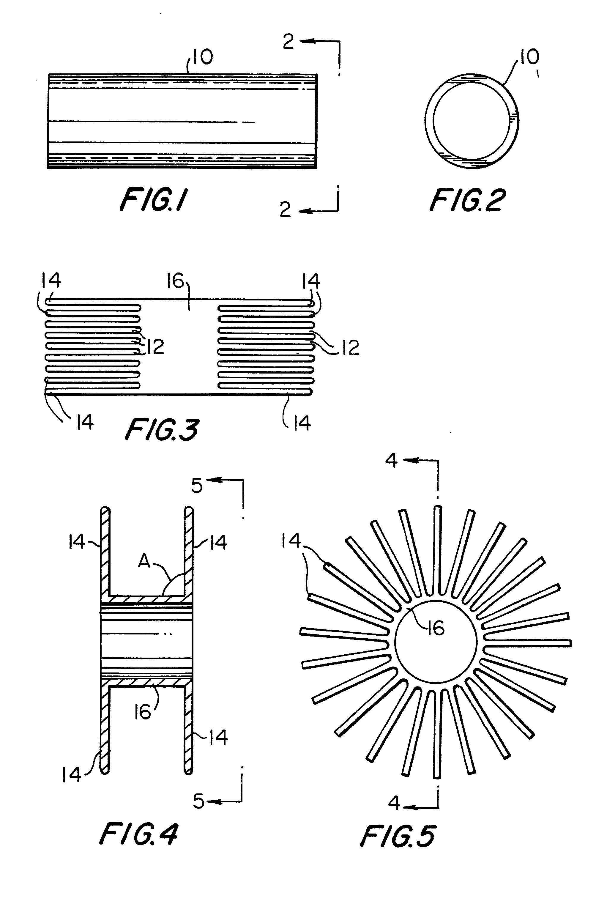
[0042]An illustrative starting component for the connector or plug structures of this invention is a hollow tube 10 as shown in FIGS. 1 and 2. Tube 10 may have any length, diameter, and wall thickness suitable for the intended use of the finished connector or plug structure. For use as a cardiac bypass graft connector, for example, tube 10 may have a diameter of about 4.0 millimeters, a wall thickness of about 0.003 inches, and a length of about 7.0 millimeters. It will be understood, however, that these specific dimensions are only exemplary, and that any other dimensions can be used instead if desired. The material of tube 10 is preferably highly elastic. A particularly preferred material is nickel titanium alloy (nitinol) metal (which can be per se conventional), but other materials such as stainless steel or thermoplastics can be used instead if desired.

[0043]A first step in processing tube 10 in accordance with the invention is to cut into it substantially axially at many locat...


PUM


Abstract

A medical graft connector or plug is made, for example, by cutting end portions of a tube of highly elastic material axially at numerous locations spaced circumferentially around the tube to produce a plurality of fingers which extend axially from each end of an uncut medial portion of the tube. The fingers are deflected radially outwardly from the medial portion and set in that condition. For a graft connector, the medial portion is coaxially connected to an end portion of a tubular graft. The connector is then installed through an aperture in the side wall of a patient's tubular body conduit, for example, by using a delivery tube in which the fingers are elastically deflected back to approximately their initial positions. When the delivery conduit is withdrawn from the connector, the fingers spring out to engage the inner and outer surfaces of the body conduit wall. For a plug, the medial portion is occluded and then the structure is installed through the aperture to be plugged in a manner similar to installation of the connector.

Description

[0001]This application is a continuation of U.S. patent application Ser. No. 09 / 569,607, filed May 10, 2000, now U.S. Pat. No. 6,660,015 which is a continuation of U.S. patent application Ser. No. 09 / 540,665, filed Mar. 31, 2000, now U.S. Pat. No. 6,391,036 which is a division of U.S. patent application Ser. No. 09 / 016,721, filed Jan. 30, 1998 now abandoned. All of these prior applications are hereby incorporated by reference herein in their entireties.BACKGROUND OF THE INVENTION[0002]This invention relates to structures that can be used to make connections between tubular medical grafts and a patient's tubular body conduits. The structures of the invention can alternatively be constructed for use as medical plugs (e.g., to close atrial or ventricular septal defects). The invention also relates to methods for making and using the structures mentioned above.[0003]Tubular grafts are frequently needed in medical procedures. For example, a coronary bypass procedure may involve the insta...

Claims


Application Information

Patent Timeline
Patent Type & Authority Patents(United States)
IPC IPC(8): A61B17/08A61B17/00A61B17/064A61B17/11A61B17/34A61F2/06
CPCA61B17/0057A61F2/064A61B17/3468A61B17/11A61B17/064A61B17/0643A61B17/08A61B17/3415A61B2017/00659A61B2017/0641
Inventor BERG, TODD ALLENPETERSON, ALEX ALDENWAHLBERG, MARK D.ST. GERMAIN, JON PATRICK
Owner ST JUDE MEDICAL ATG
Why Eureka
Try Eureka