
Display method and apparatus of x-ray projection image for medical use, x-ray CT apparatus for medical use and recording medium for recording program to achieve the display method

Inactive Publication Date: 2006-05-02
NIHON UNIVERSITY +1
View PDF1 Cites 39 Cited by
  • Summary
  • Abstract
  • Description
  • Claims
  • Application Information

AI Technical Summary

Benefits of technology

[0009]In such a manner, three dimensional X-ray absorption coefficient data existing in a direction substantially along the X-ray radiating direction so that clear images can be obtained. In addition, three dimensional X-ray absorption coefficient data are projected on the flat projection plane. Therefore, exemplifying a dental arch, images seen from a projecting direction perpendicular to the projection plane can be obtained and they are easily comprehensive images by intuition for dental diagnosis. Further, perspectively observable image like the dental arch, namely an object to be examined, is turned by sequentially obtaining and displaying X-ray projection images by rotating a direction in which the X-ray projection image is to be projected, that is the projection plane, thus obtaining highly comprehensive and highly advantageous images for diagnosis.

Problems solved by technology

Further, the panoramic images were generally used images for diagnosis in the dental field, however they weren't easily understood by intuition as a dental arch was actually seen from one direction.
Although the condition of a dental root could be understood by the obtained X-ray panoramic images, it was difficult to distinguish where the root exists from a cheek to a tongue.

Method used


Image

  • Display method and apparatus of x-ray projection image for medical use, x-ray CT apparatus for medical use and recording medium for recording program to achieve the display method
  • Display method and apparatus of x-ray projection image for medical use, x-ray CT apparatus for medical use and recording medium for recording program to achieve the display method
  • Display method and apparatus of x-ray projection image for medical use, x-ray CT apparatus for medical use and recording medium for recording program to achieve the display method

Examples


Embodiment Construction

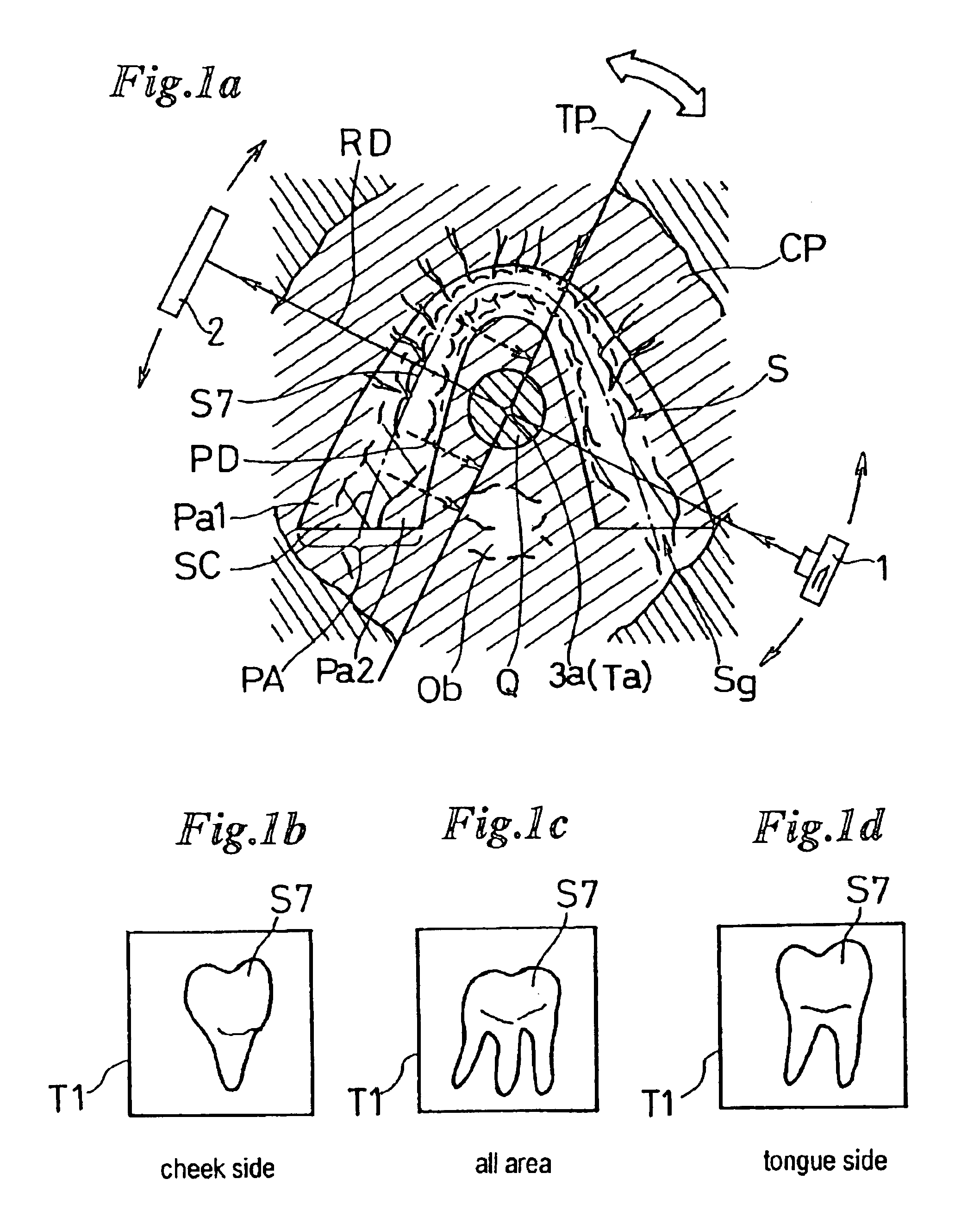
[0039]FIG. 1a is a conceptual diagram explaining a procedure of a display method of X-ray projection images for medical use according to the present invention, FIGS. 1b, 1c and 1d show an example of X-ray projection images of a tooth shown according to the method.

[0040]FIG. 1a shows an image wherein three dimensional X-ray absorption coefficient data obtained by an X-ray CT method in which an X-ray generator 1 and an X-ray detector 2 are opposed and rotated around a dental arch S, being an object to be examined, are seen from an image layer CP having a predetermined thickness in a direction perpendicular to a radiated plane obtained by rotation of X-ray radiation, namely shows an image when three dimensional X-ray absorption coefficient data are seen from an axial direction of a rotation center 3a of X-ray radiation.

[0041]Circular area around the center of the image layer CP is called as a virtual local region Q which is always locally radiated with X-rays in a radiography wherein a...


PUM


Abstract

A display method and apparatus of an X-ray projection image for medical use wherein a projection interested area to construct the X-ray projection image of an object is set on an image layer with an extending predetermined thickness perpendicular to an X-ray radiation plane defined by rotational radiation of an X-ray beam, the projection interested area is comprised of the three dimensional X-ray absorption coefficient data, and the X-ray projection image is produced by projecting the three dimensional X-ray absorption coefficient data existing in the projection interested area with respect to a projection plane intersecting the X-ray radiation plane.

Description

TECHNICAL FIELD[0001]The present invention relates to a display method of an X-ray projection image for medical use based on three dimensional X-ray absorption coefficient data obtained by an X-ray computed tomography (called CT hereinafter) method in which an X-ray image of an object to be examined is obtained by turning an X-ray generator and an X-ray detector faced each other, to its display apparatus, to an X-ray CT apparatus using the display method and a recording medium for recording a program to achieve the display method.BACKGROUND ART[0002]The applicants have proposed an X-ray CT method and apparatus in which conical X-ray beams with remarkably small sectional radiation area are locally radiated only on a local region, a part of an object, to obtain clear three dimensional X-ray absorption coefficient data on the region. For example, JP-A-2000-139902 discloses its CT method and apparatus.[0003]In this laid open publication, the applicants have proposed a basic X-ray CT met...

Claims


Application Information

Patent Timeline
IPC IPC(8): A61B6/14
CPCA61B6/0478A61B6/469A61B6/463A61B6/14A61B6/51
Inventor ARAI, YOSHINORISUZUKIYASUDA, KOUJI
Owner NIHON UNIVERSITY
Features
  • Generate Ideas
  • Intellectual Property
  • Life Sciences
  • Materials
  • Tech Scout
  • Unparalleled Data Quality
  • Higher Quality Content
  • 60% Fewer Hallucinations
Learn More